[go: up one dir, main page]
More Web Proxy on the site http://driver.im/
You seem to have javascript disabled. Please note that many of the page functionalities won't work as expected without javascript enabled.


Feature Review Papers for Life

A topical collection in Life (ISSN 2075-1729).

Viewed by 279522

Editors


E-Mail Website
Guest Editor
1. Department of Chemistry, University of Turin, 10125 Turin, Italy
2. Jet Propulsion Laboratory, California Institute of Technology, Pasadena, CA 91125, USA
Interests: emergence of life; emergence of oxygenic photosynthesis; giant sedimentary exhalative orebodies; nanoengines; chemobrionics

E-Mail Website
Guest Editor
Astrobiology Group, Center for Astronomy and Astrophysics, Technical University Berlin, Berlin, Germany
Interests: planetary habitability; astrobiology; evolutionary biology; extreme environments; geobiology; space missions
Special Issues, Collections and Topics in MDPI journals

E-Mail Website
Guest Editor
Department of Bioinformatics, Kyushu Institute of Technology, Fukuoka 804-8550, Japan
Interests: plant genomics; plant evolution; system biology in plants
Special Issues, Collections and Topics in MDPI journals

E-Mail Website
Guest Editor
Division of Nephrology and Hypertension, 1st Department of Internal Medicine, Medical School, AHEPA Hospital, Aristotle University, 57001 Thessaloniki, Greece
Interests: CKD; hemodialysis; oxidative stress; vitamin K; diabetic nephropathy; vascular calcification; CVD; matrix GLA protein
Special Issues, Collections and Topics in MDPI journals

E-Mail Website
Guest Editor
The Second Department of Internal Medicine, Osaka Medical College, 2-7, Daigakucho, Takatsukishi, Osaka 569-8686, Japan
Interests: liver cirrhosis; sarcopenia; liver-gut axis; viral hepatitis; hepatocellular carcinoma
Special Issues, Collections and Topics in MDPI journals

Topical Collection Information

Dear Colleagues,

This Special Issue aims to collect high quality review papers in the Life sciences’ research fields. We encourage researchers from various fields within the journal’s scope to contribute review papers highlighting the latest developments in their research field, or to invite relevant experts and colleagues to do so.  The topic of this Special Issue includes, but is not limited to:

origins of life, astrobiology, biology, protein, ecology, genetics, plant science, animal science, medicine, physiology and pathology.

Such review papers should provide syntheses of ideas and have the potential to challenge existing paradigms and create new frameworks that will advance our understanding of all aspect of Life.

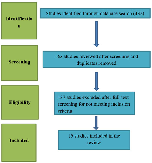
Review manuscripts should comprise the front matter, literature review sections and the back matter. The template file can also be used to prepare the front and back matter of your review manuscript. It is not necessary to follow the remaining structure. Structured reviews and meta-analyses should use the same structure as research articles and ensure they conform to the PRISMA guidelines.

Prof. Dr. Michael Russell
Prof. Dr. Dirk Schulze-Makuch
Prof. Dr. Kousuke Hanada
Dr. Stefanos Roumeliotis 
Dr. Hiroki Nishikawa

Dr. Alfredo Conti
Guest Editors

Manuscript Submission Information

Manuscripts should be submitted online at www.mdpi.com by registering and logging in to this website. Once you are registered, click here to go to the submission form. Manuscripts can be submitted until the deadline. All submissions that pass pre-check are peer-reviewed. Accepted papers will be published continuously in the journal (as soon as accepted) and will be listed together on the collection website. Research articles, review articles as well as short communications are invited. For planned papers, a title and short abstract (about 100 words) can be sent to the Editorial Office for announcement on this website.

Submitted manuscripts should not have been published previously, nor be under consideration for publication elsewhere (except conference proceedings papers). All manuscripts are thoroughly refereed through a single-blind peer-review process. A guide for authors and other relevant information for submission of manuscripts is available on the Instructions for Authors page. Life is an international peer-reviewed open access monthly journal published by MDPI.

Please visit the Instructions for Authors page before submitting a manuscript. The Article Processing Charge (APC) for publication in this open access journal is 2600 CHF (Swiss Francs). Submitted papers should be well formatted and use good English. Authors may use MDPI's English editing service prior to publication or during author revisions.

Published Papers (60 papers)

2024

Jump to: 2023, 2022, 2021

44 pages, 2974 KiB  
Review
Exploring Aquaporins in Human Studies: Mechanisms and Therapeutic Potential in Critical Illness
by Charikleia S. Vrettou, Vasileios Issaris, Stelios Kokkoris, Georgios Poupouzas, Chrysi Keskinidou, Nikolaos S. Lotsios, Anastasia Kotanidou, Stylianos E. Orfanos, Ioanna Dimopoulou and Alice G. Vassiliou
Life 2024, 14(12), 1688; https://doi.org/10.3390/life14121688 - 20 Dec 2024
Viewed by 492
Abstract
Aquaporins (AQPs) are membrane proteins facilitating water and other small solutes to be transported across cell membranes. They are crucial in maintaining cellular homeostasis by regulating water permeability in various tissues. Moreover, they regulate cell migration, signaling pathways, inflammation, tumor growth, and metastasis. [...] Read more.
Aquaporins (AQPs) are membrane proteins facilitating water and other small solutes to be transported across cell membranes. They are crucial in maintaining cellular homeostasis by regulating water permeability in various tissues. Moreover, they regulate cell migration, signaling pathways, inflammation, tumor growth, and metastasis. In critically ill patients, such as trauma, sepsis, and patients with acute respiratory distress syndrome (ARDS), which are frequently encountered in intensive care units (ICUs), water transport regulation is crucial for maintaining homeostasis, as dysregulation can lead to edema or dehydration, with the latter also implicating hemodynamic compromise. Indeed, AQPs are involved in fluid transport in various organs, including the lungs, kidneys, and brain, where their dysfunction can exacerbate conditions like ARDS, acute kidney injury (AKI), or cerebral edema. In this review, we discuss the implication of AQPs in the clinical entities frequently encountered in ICUs, such as systemic inflammation and sepsis, ARDS, AKI, and brain edema due to different types of primary brain injury from a clinical perspective. Current and possible future therapeutic implications are also considered. Full article
Show Figures

Figure 1

Figure 1
<p>Aquaporin structure. (<b>a</b>) AQP side monomer view parallel to the membrane (the two NPA motifs are shown in yellow), (<b>b</b>) top view of the AQP tetramer from the extracellular side (water molecules in the pore are shown as red spheres), (<b>c</b>) the AQP monomer with the six transmembrane alpha-helices (1–6) connected by five loops extending into the extracellular and cytoplasmic spaces (A–E), and (<b>d</b>) illustration of the AQP hourglass-shaped channel, depicting the two selectivity filters (ar/R and NPA), allowing the passage of water molecules and other solutes, as shown, while excluding ions and larger solutes. Abbreviations: AQP, aquaporin; ar/R, aromatic/arginine; NPA, asparagine-proline-alanine. Image adapted from [<a href="#B33-life-14-01688" class="html-bibr">33</a>,<a href="#B83-life-14-01688" class="html-bibr">83</a>]. Under Creative Commons CC BY 4.0 license.</p>
Full article ">Figure 2
<p>Regulation of aquaporin expression and function. Aquaporin gene expression is tightly regulated by several factors including cytokine levels, hypoxia, cell osmotic conditions, and vasopressin. Following RNA expression, alternative splicing events and regulatory molecules such as micro RNAs (miRNAs), act upon the RNA molecules, thus regulating protein production. A series of post-translational modifications, including phosphorylation, methylation, and ubiquitination determine the function and localization of the formed proteins. AQPs may be alternatively stored in intracellular vesicles to be rapidly transported to the cell membrane upon stimulation. Abbreviations: AQP, aquaporin; miRNAs, micro RNAs.</p>
Full article ">Figure 3
<p>Aquaporin expression in human immune cells and their proposed roles in sepsis. AQP1 is expressed in neutrophils, monocytes, and lymphocytes, contributing to water transport and cell volume regulation during migration and differentiation. AQP3 is expressed in macrophages, lymphocytes, and dendritic cells, facilitating chemokine-driven migration and supporting energy metabolism through glycerol transport. AQP5 is expressed in some immune cells, particularly lymphocytes, neutrophils, and dendritic cells. AQP5 plays a role in facilitating water transport, which is critical for maintaining cell shape and motility during migration. AQP9 is most prominently expressed in leukocytes, aiding in respiratory bursts and cell motility during pathogen defense. It is expressed in neutrophils, macrophages, monocytes, lymphocytes, and dendritic cells. Other AQPs detected at low concentrations in immune cells are AQP6, AQP7, AQP8, AQP11, and AQP12, expressed in monocytes, macrophages, and dendritic cells. AQPs are important regulators of fluid balance/edema, inflammation/immune response, and vascular permeability. During inflammatory conditions, such as sepsis, AQP expression is altered and correlates with disease severity and outcomes. Abbreviations: AQP, aquaporin; ROS, reactive oxygen species. Image adapted from [<a href="#B117-life-14-01688" class="html-bibr">117</a>].</p>
Full article ">Figure 4
<p>Aquaporins in the human lung. The lung facilitates oxygen and carbon dioxide exchange between air and blood. Maintaining the fluid volume is essential for gas exchange, achieved by balancing fluid transport across the epithelium. Aquaporins (AQPs) are integral membrane proteins that act as water channels, facilitating water transport across cell membranes in response to osmotic gradients. Under physiological conditions, aquaporins usually facilitate fluid management by transporting it out of the alveolar space into the interstitium or capillaries. Τhe main aquaporins expressed in the lung are AQP1, AQP3, AQP4, and AQP5. AQP1 is present in the capillary endothelial cells and controls water movement across the endothelial barrier. AQP3 and AQP4 are found in the airway epithelium and alveolar epithelial cells. AQP5 is expressed in the alveolar epithelial cells type I and is primarily responsible for water transport across the alveolar barrier. Acute respiratory distress syndrome (ARDS) constitutes a severe lung condition characterized by the widespread release of pro- and anti-inflammatory cytokines, oxidative stress (production of reactive oxygen species-ROS), increased vascular permeability, and accumulation of fluid in the alveoli, which leads to impaired gas exchange and respiratory failure. In ARDS, increased water transport by AQPs could contribute to fluid accumulation in the alveoli if the inflammation and barrier dysfunction are uncontrolled, exacerbating pulmonary edema. Disruption of the expression or function of these AQPs, as is often observed in ARDS, compromises the lung’s ability to clear fluid, leading to worsened pulmonary edema. Abbreviations: AQP, aquaporin; ROS, reactive oxygen species. Adapted from [<a href="#B157-life-14-01688" class="html-bibr">157</a>].</p>
Full article ">Figure 5
<p>Aquaporins in the human kidney. Aquaporins (AQPs) play a critical role in kidney function by regulating water transport across membranes, crucial for urine concentration and maintaining fluid balance. AQP1 facilitates water reabsorption in the proximal tubules and descending limb of the loop of Henle, while AQP2 is key in the collecting ducts, regulated by antidiuretic hormone (ADH) to concentrate urine. AQPs 3 and 4 support water exit from the collecting duct cells to the bloodstream. AQP7 is expressed in the brush border of the S3 segment of the proximal tubule and plays a significant role in metabolism by regulating glycerol transport. AQP8 enables bidirectional water and hydrogen peroxide transport across biological membranes, while AQP11 is uniquely located in the endoplasmic reticulum membrane of proximal tubular cells. Dysfunction of AQPs can lead to conditions like nephrogenic diabetes insipidus or fluid imbalances. Panels on the left show proposed mechanisms of aquaporin involvement in AKI. The panel on the right shows approaches that could regulate AQP expression. The downward arrows designate decreased expression. Abbreviations: AKI, acute kidney injury; AQP, aquaporin; H<sub>2</sub>O<sub>2</sub>, hydrogen peroxide; I/R, ischemia/reperfusion; LPS, lipopolysaccharide. Image reproduced from [<a href="#B24-life-14-01688" class="html-bibr">24</a>]. Aspects of this image were partially adapted. Under Creative Commons CC BY 4.0 license.</p>
Full article ">Figure 6
<p>Main pathophysiological roles of aquaporins in acute brain injury. Aquaporins (AQPs) are pivotal in the pathophysiology of acute brain injury, and their role extends across key processes such as cerebral edema formation, blood–brain barrier regulation, and inflammatory response modulation. AQP4 has emerged as a central player, influencing water homeostasis and edema resolution, while other aquaporins, such as AQP1, AQP2, and AQP9, contribute to diverse pathophysiological mechanisms, including immune cell migration and metabolic adaptation. Aspects of this figure were adapted with permission from Servier Medical Art library, available under Creative Commons license.</p>
Full article ">
16 pages, 812 KiB  
Review
Mechanisms of Cbl-Mediated Ubiquitination of Proteins in T and Natural Killer Cells and Effects on Immune Cell Functions
by Pulak Ranjan Nath and Noah Isakov
Life 2024, 14(12), 1592; https://doi.org/10.3390/life14121592 - 3 Dec 2024
Viewed by 676
Abstract
Post-translational ubiquitination is an essential mechanism for the regulation of protein stability and function, which contributes to the regulation of the immune system. Cbl, an E3 ubiquitin ligase, is particularly well-characterized in the context of T and NK cell signaling, where it serves [...] Read more.
Post-translational ubiquitination is an essential mechanism for the regulation of protein stability and function, which contributes to the regulation of the immune system. Cbl, an E3 ubiquitin ligase, is particularly well-characterized in the context of T and NK cell signaling, where it serves as a key regulator of receptor downstream signaling events and as a modulator of cell activation. Cbl promotes the proteasomal degradation of TCR/CD3 subunits as well as the protein kinases Fyn and Lck in T cells. Additionally, the scaffold protein linker for activation of T cells (LAT) is a universal target for Cbl-mediated ubiquitination and degradation in both T and NK cells. Recent findings suggest that CrkII-mediated ubiquitination and degradation of C3G by Cbl during early T cell activation may also be relevant to NK cell signaling. Given its role in modulating immune responses and its manageable impact on autoimmunity, Cbl is being investigated as a target for cancer immunotherapy. This review explores the ubiquitin ligase activity of Cbl and its implications for CAR T and NK cell immunotherapies. Full article
Show Figures

Figure 1

Figure 1
<p>Schematic structure of Cbl-b and mechanism of action. The upper figure shows the schematic structure of Cbl-b protein. The N-terminal tyrosine kinase binding domain (TKBD) is composed of a four-helical bundle, a calcium-binding EF-hand, and an SH2 domain. A short helical region links the N-terminal TKBD and C-terminal RING finger domain, followed by a polyproline-rich region (PPP). The lower figure models the Cbl ubiquitin ligase function. SH2 domain (green) and PPP-regions of Cbl-b bind to phospho-tyrosine (pTyr) and SH3-domain-bearing proteins, respectively. Ubiquitin (UB, orange) is activated by E1 ligase and transferred to a ubiquitin-conjugating enzyme (E2 ligase, pink). E2 ligase interacts with Cbl-b via its RING domain (blue) and brings UB to the proximity of Cbl-b-interacting proteins and promotes their mono- or polyubiquitination. The figure was created using BioRender.com (<a href="http://www.biorender.com" target="_blank">www.biorender.com</a>, accessed on 24 November 2024).</p>
Full article ">Figure 2
<p>Regulation of signal transduction pathways and function of T cells by Cbl. Upon binding to the cognate peptidyl-MHC complex and stabilization by the CD4/CD8 co-receptor, the T cell receptor (TCR)/CD3 complex initiates a number of downstream activation signals that eventually determine cell fate through regulating cytokine production, cell survival, proliferation, and differentiation. An early event in TCR activation is phosphorylation of immunoreceptor tyrosine-based activation motifs (ITAMs) on the cytosolic side of the TCR/CD3 complex by lymphocyte protein tyrosine kinase (Lck). The CD45 receptor tyrosine phosphatase modulates the phosphorylation and activation of Lck that is brought by the CD4 or CD8 co-receptor to the proximity of the TCR/CD3 complex. Zeta-chain-associated protein kinase (Zap-70) is recruited to the TCR/CD3 complex, where it becomes activated, promoting recruitment and phosphorylation of downstream adaptor or scaffold proteins, like CT10 regulator of kinase (Crk). Crk recruits C3G (a guanine nucleotide exchange factor) that in turn activates the Ras/Rap1 signaling pathways, the MAPK/Erk pathways, and the AP1 transcription factor. ZAP-70 also phosphorylates the scaffold protein, Linker for activation of T cells (LAT), which recruits phospholipase C γ1 (PLCγ1), which hydrolyzes phosphatidylinositol 4,5-bisphosphate (PIP2) to form the diacylglycerol (DAG) and inositol trisphosphate (IP3) second messengers. DAG activates PKCθ and the MAPK/Erk pathways, both promoting transcription factor NF-κB. IP3 triggers the release of Ca2+ from the ER, which promotes the entry of extracellular Ca2+ into cells through calcium release-activated Ca2+ (CRAC) channels. Calcium-bound calmodulin (Ca2+/CaM) activates the phosphatase calcineurin, which promotes IL-2 gene transcription through the transcription factor NFAT. One of the effects of the Cbl-b protein in T cells is to downregulate the CD3- and CD28-linked signal transduction pathways. Cbl-b can ubiquitinate the T cell antigen receptor (TCR) CD3ζ chain and the LAT adapter protein and direct them to proteasomal degradation. In resting T cells, the CrkII adapter protein constitutively associates with the guanine nucleotide exchange factor C3G and maintains a basal level of signaling that is insufficient for cell activation. However, upon TCR stimulation, CrkII also interacts with Cbl-b (via a distinct protein–protein binding site) and brings Cbl-b into close proximity with C3G to enable the ubiquitination of C3G by Cbl-b. The ubiquitinated C3G undergoes proteasomal degradation, thereby promoting signal termination and preventing persistent immune cell activation. The figure was created using BioRender.com (<a href="http://www.biorender.com" target="_blank">www.biorender.com</a>, accessed on 14 July 2024).</p>
Full article ">Figure 3
<p>Regulation of signal transduction pathways and function of NK cells by Cbl. c-Cbl ubiquitinates the stimulatory receptor NKG2D on NK cells, leading to the endocytosis and degradation of the activation signaling complex. c-Cbl also mediates non-degradative ubiquitination of VAV1, a regulator of signal transduction pathways downstream of the NKG2D and 2B4 stimulatory receptors, and negatively regulates their downstream signaling pathways in NK cells. Upon interaction with the cognitive ligands, the growth arrest-specific gene 6 (Gas6) and protein S (ProS) and the Tyro3, Axl, and Mer (TAM)-family receptor tyrosine kinases (RTKs) phosphorylate and activate Cbl-b, which in turn promotes the ubiquitination of the adapter protein linker for activation of T cells (LAT), followed by its proteasomal degradation. This results in the inhibition of LAT-dependent signaling pathways that otherwise promote cytokine and chemokine production in NK cells. The figure was created using BioRender.com (<a href="http://www.biorender.com" target="_blank">www.biorender.com</a>, accessed on 14 July 2024).</p>
Full article ">
13 pages, 532 KiB  
Review
Criteria for Assessing Exposure to Biomechanical Risk Factors: A Research-to-Practice Guide—Part 1: General Issues and Manual Material Handling
by Francesca Graziosi, Roberta Bonfiglioli, Francesco Decataldo and Francesco Saverio Violante
Life 2024, 14(11), 1398; https://doi.org/10.3390/life14111398 - 30 Oct 2024
Viewed by 862
Abstract
Musculoskeletal disorders are the most prevalent occupational health problem all over the world and are often related to biomechanical risk factors; to control these risk factors, several assessment methods (mostly observational) have been proposed in the past 40 years. An in-depth knowledge of [...] Read more.
Musculoskeletal disorders are the most prevalent occupational health problem all over the world and are often related to biomechanical risk factors; to control these risk factors, several assessment methods (mostly observational) have been proposed in the past 40 years. An in-depth knowledge of each method to evaluate biomechanical risk factors is needed to effectively employ them in the field, together with a robust understanding of their effective predictive value and limitations. In Part 1, some general issues relevant to biomechanical risk assessment are discussed, and the method for assessing manual material handling after receiving more robust validation data is reviewed (Revised NIOSH Lifting Equation), together with a discussion about variability of tasks. Similarly, for the assessment of the biomechanical exposure of the upper limb, the TLV for Hand activity (ACGIH®) is presented in Part 2 of this guide, together with criteria to proportion risk assessment to the working duration in part-time jobs. Full article
Show Figures

Figure 1

Figure 1
<p>Schematic representation of the biomechanical risk assessment evaluation procedure for manual material handling.</p>
Full article ">
18 pages, 1514 KiB  
Review
Clinician’s Guide to Epitranscriptomics: An Example of N1-Methyladenosine (m1A) RNA Modification and Cancer
by Ana Kvolik Pavić, Josipa Čonkaš, Ivan Mumlek, Vedran Zubčić and Petar Ozretić
Life 2024, 14(10), 1230; https://doi.org/10.3390/life14101230 - 25 Sep 2024
Viewed by 1339
Abstract
Epitranscriptomics is the study of modifications of RNA molecules by small molecular residues, such as the methyl (-CH3) group. These modifications are inheritable and reversible. A specific group of enzymes called “writers” introduces the change to the RNA; “erasers” delete it, [...] Read more.
Epitranscriptomics is the study of modifications of RNA molecules by small molecular residues, such as the methyl (-CH3) group. These modifications are inheritable and reversible. A specific group of enzymes called “writers” introduces the change to the RNA; “erasers” delete it, while “readers” stimulate a downstream effect. Epitranscriptomic changes are present in every type of organism from single-celled ones to plants and animals and are a key to normal development as well as pathologic processes. Oncology is a fast-paced field, where a better understanding of tumor biology and (epi)genetics is necessary to provide new therapeutic targets and better clinical outcomes. Recently, changes to the epitranscriptome have been shown to be drivers of tumorigenesis, biomarkers, and means of predicting outcomes, as well as potential therapeutic targets. In this review, we aimed to give a concise overview of epitranscriptomics in the context of neoplastic disease with a focus on N1-methyladenosine (m1A) modification, in layman’s terms, to bring closer this omics to clinicians and their future clinical practice. Full article
Show Figures

Graphical abstract

Graphical abstract
Full article ">Figure 1
<p>Comparative molecular structures of unmodified RNA nucleosides and ribose with some of the most common RNA modifications. Created with <a href="https://BioRender.com" target="_blank">https://BioRender.com</a> (accessed on 16 May 2024).</p>
Full article ">Figure 2
<p>Location of m<sup>1</sup>A modifications within the cloverleaf structure of tRNA molecule (in red). D-loop (in purple) is named after dihydrouridine (D or DHU), a modified nucleotide generally present in this region. The anticodon loop contains the anticodon (in dark green), which recognizes and binds to a specific codon on mRNA during protein translation. Variable loop (in magenta) varies from 3–21 bases and is used for classification of tRNAs. T-loop or TΨC loop (in light green) contains modified uridine, a pseudouridine (Ψ). Acceptor stem is formed by the base pairing of the 5′ end and the 3′ end of tRNA. Amino acid binding site or CCA tail (in blue) is a cytosine–cytosine–adenine motif at the 3′ end of tRNA, and amino acid is covalently bonded to it by aminoacyl tRNA synthetase. Created with <a href="https://BioRender.com" target="_blank">https://BioRender.com</a> (accessed on 28 June 2024).</p>
Full article ">Figure 3
<p>Some of the known readers (green), writers (blue), and erasers (ochre) of the m<sup>1</sup>A modification of the tRNA molecule. Created with <a href="https://BioRender.com" target="_blank">https://BioRender.com</a> (accessed on 17 May 2024).</p>
Full article ">
10 pages, 275 KiB  
Review
Metabolic Shift and Hyperosmolarity Underlie Age-Related Macular Degeneration
by Laurent Schwartz, Jules Schwartz, Marc Henry and Ashraf Bakkar
Life 2024, 14(9), 1189; https://doi.org/10.3390/life14091189 - 20 Sep 2024
Viewed by 1235
Abstract
Age-related macular degeneration (AMD) is both a poorly understood and devastating disease. Here, we analyze the physico-chemical forces at stake, including osmolarity, redox shift, and pressure due to inflammation. Hyperosmolarity plays a key role in diseases of the anterior segment of the eye [...] Read more.
Age-related macular degeneration (AMD) is both a poorly understood and devastating disease. Here, we analyze the physico-chemical forces at stake, including osmolarity, redox shift, and pressure due to inflammation. Hyperosmolarity plays a key role in diseases of the anterior segment of the eye such as glaucoma, cataracts or dry eyes, and corneal ulceration. However, its role in macular degeneration has been largely overlooked. Hyperosmolarity is responsible for metabolic shifts such as aerobic glycolysis which increases lactate secretion by Muller cells. Increased osmolarity will also cause neoangiogenesis and cell death. Because of its unique energetic demands, the macula is very sensitive to metabolic shifts. As a proof of concept, subretinal injection of drugs increasing hyperosmolarity such as polyethylene glycol causes neoangiogenesis and drusen-like structures in rodents. The link between AMD and hyperosmolarity is reinforced by the fact that treatments aiming to restore mitochondrial activity, such as lipoic acid and/or methylene blue, have been experimentally shown to be effective. We suggest that metabolic shift, inflammation, and hyperosmolarity are hallmarks in the pathogenesis and treatment of AMD. Full article
Show Figures

Figure 1

Figure 1
<p>Correlation between inflammation, hyperosmolarity, and metabolic shift in AMD.</p>
Full article ">
19 pages, 1537 KiB  
Review
Understanding Galectin-3’s Role in Diastolic Dysfunction: A Contemporary Perspective
by Wen-Rui Hao, Chun-Han Cheng, Ju-Chi Liu, Huan-Yuan Chen, Jin-Jer Chen and Tzu-Hurng Cheng
Life 2024, 14(7), 906; https://doi.org/10.3390/life14070906 - 20 Jul 2024
Cited by 2 | Viewed by 1461
Abstract
Diastolic dysfunction, a prevalent condition characterized by impaired relaxation and filling of the left ventricle, significantly contributes to heart failure with preserved ejection fraction (HFpEF). Galectin-3, a β-galactoside-binding lectin, has garnered attention as a potential biomarker and mediator of fibrosis and inflammation in [...] Read more.
Diastolic dysfunction, a prevalent condition characterized by impaired relaxation and filling of the left ventricle, significantly contributes to heart failure with preserved ejection fraction (HFpEF). Galectin-3, a β-galactoside-binding lectin, has garnered attention as a potential biomarker and mediator of fibrosis and inflammation in cardiovascular diseases. This comprehensive review investigates the impact of galectin-3 on diastolic dysfunction. We explore its molecular mechanisms, including its involvement in cellular signaling pathways and interaction with components of the extracellular matrix. Evidence from both animal models and clinical studies elucidates galectin-3’s role in cardiac remodeling, inflammation, and fibrosis, shedding light on the underlying pathophysiology of diastolic dysfunction. Additionally, we examine the diagnostic and therapeutic implications of galectin-3 in diastolic dysfunction, emphasizing its potential as both a biomarker and a therapeutic target. This review underscores the significance of comprehending galectin-3’s role in diastolic dysfunction and its promise in enhancing diagnosis and treatment approaches for HFpEF patients. Full article
Show Figures

Figure 1

Figure 1
<p>Cellular signaling pathways of galectin-3 in cardiac diastolic pathophysiology.</p>
Full article ">Figure 2
<p>The pathophysiology of galectin-3 interacted with extracellular matrix components in cardiac diastolic dysfunction.</p>
Full article ">Figure 3
<p>The role of galectin-3 in inflammation, fibrosis, and heart remodeling leading to cardiac diastolic dysfunction.</p>
Full article ">
10 pages, 846 KiB  
Review
The Current Role of Contrast-Enhanced Ultrasound (CEUS) in the Diagnosis and Staging of Bladder Cancer: A Review of the Available Literature
by Valerio Santarelli, Davide Rosati, Vittorio Canale, Stefano Salciccia, Giovanni Di Lascio, Giulio Bevilacqua, Antonio Tufano, Alessandro Sciarra, Vito Cantisani, Giorgio Franco, Martina Moriconi and Giovanni Battista Di Pierro
Life 2024, 14(7), 857; https://doi.org/10.3390/life14070857 - 9 Jul 2024
Viewed by 1767
Abstract
Contrast-enhanced ultrasound (CEUS) is an advanced imaging technique that integrates conventional US with the intravenous injection of specific US contrast agents (UCAs), combining the non-invasiveness of US with the higher accuracy of contrast-enhanced imaging. In contrast with magnetic resonance imaging (MRI), computed tomography [...] Read more.
Contrast-enhanced ultrasound (CEUS) is an advanced imaging technique that integrates conventional US with the intravenous injection of specific US contrast agents (UCAs), combining the non-invasiveness of US with the higher accuracy of contrast-enhanced imaging. In contrast with magnetic resonance imaging (MRI), computed tomography (CT) and cystoscopy, CEUS has few contraindications, and UCAs are non-nephrotoxic agents that can be safely used in patients with kidney failure. CEUS is a well-established method for the detection of liver lesions and for echocardiography, and its indications are expanding. The updated 2018 WFUMB-EFSUMB guidelines have added the urinary bladder under non-hepatic applications of CEUS. The technique is able to distinguish between benign tissue, such as clots or hematoma, and malignant lesions by perfusing the mass with contrast agent. Thanks to the different perfusion rates of the various layers of the bladder wall, CEUS is also able to predict tumor invasion depth and stage. Despite that, current urological guidelines do not include CEUS as a plausible imaging technique for bladder urothelial carcinoma. The main reason for this omission might be the presence of scarce randomized evidence and the absence of large validated series. In this review, we describe the rationale behind the use of CEUS in bladder cancer and the added value of this imaging technique in the detection and staging of bladder lesions. In addition, we researched the available literature on the topic and then described the results of randomized clinical trials and a meta-analysis investigating the accuracy of CEUS in bladder cancer diagnosis and staging. The reported studies show that CEUS is a highly accurate diagnostic and staging tool for BC, reaching levels of specificity and sensitivity in differentiating between Ta-T1, or low-grade BC, and T2, or high-grade BC, that are comparable to those shown by the reference standard methods. Nonetheless, several limitations were found and are highlighted in this review. The aim of this study is to further validate and promote the use of CEUS as a quick, economic and effective diagnostic tool for this high-impact disease. Full article
Show Figures

Figure 1

Figure 1
<p>CEUS performance. Red box: Normal bladder wall; orange box: NMIBC; blue box: MIBC. UCA, ultrasound contrast agent; RT, rise time; PI, peak intensity. TICs (time intensity curves): curve A, normal bladder wall; curve B, low-grade carcinoma; curve C, high-grade carcinoma.</p>
Full article ">Figure 2
<p>Comparison between greyscale US, CEUS and CDUS regarding the appearance of a solid tumor (indicated by a blue arrow). (<b>A</b>) On greyscale US, the tumor appears as an echogenic protuberance of the bladder wall. (<b>B</b>) The same mass shows a strong early enhancement after SonoVue injection, highly suggestive of malignancy. (<b>C</b>) CDUS shows vascularity mostly to the base of the lesion, raising suspicion for malignancy but less clearly than CEUS [<a href="#B34-life-14-00857" class="html-bibr">34</a>].</p>
Full article ">
15 pages, 1275 KiB  
Review
Combination of H1 and H2 Histamine Receptor Antagonists: Current Knowledge and Perspectives of a Classic Treatment Strategy
by Erwen Kou, Xiaobei Zhang, Baiping Dong, Bo Wang and Yuanjie Zhu
Life 2024, 14(2), 164; https://doi.org/10.3390/life14020164 - 23 Jan 2024
Cited by 2 | Viewed by 14171
Abstract
Histamine receptor antagonists, which can bind to specific histamine receptors on target cells, exhibit substantial therapeutic efficacy in managing a variety of histamine-mediated disorders. Notably, histamine H1 and H2 receptor antagonists have been extensively investigated and universally acknowledged as recommended treatment agents for [...] Read more.
Histamine receptor antagonists, which can bind to specific histamine receptors on target cells, exhibit substantial therapeutic efficacy in managing a variety of histamine-mediated disorders. Notably, histamine H1 and H2 receptor antagonists have been extensively investigated and universally acknowledged as recommended treatment agents for numerous allergic diseases and acid-related disorders, respectively. Historically, the combination of H1 and H2 receptor antagonists has been considered a classic treatment strategy, demonstrating relatively superior efficacy compared with single-drug therapies in the treatment of diverse histamine-mediated diseases. The latest emerging studies have additionally suggested the underlying roles of histamine and H1R and H2R in the development of anxiety disorders, arthritic diseases, and postexercise hypotension. Nevertheless, there is still a lack of systematic reviews on the clinical efficacy of combination therapy, greatly limiting our understanding of its clinical application. Here, we present a comprehensive overview of the current knowledge and perspectives regarding the combination of H1 and H2 histamine receptor antagonists in various histamine-mediated disorders. Furthermore, we critically analyze the adverse effects and limitations associated with combination therapy while suggesting potential solutions. Our review can offer a systematic summary and promising insights into the in-depth and effective application of the combination of H1 and H2 receptor antagonists. Full article
Show Figures

Figure 1

Figure 1
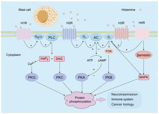
<p>Schematic overview of the main signaling pathways by which histamine exerts its biological effects on target cells. Presently, four subtypes of histamine receptors have been identified, namely, H1R, H2R, H3R, and H4R. H1R exerts its effects mainly by coupling to Gq/11 proteins and subsequently elicits the activation of phospholipase C. This lipase can produce 1,2-diacylglycerol and inositol-1,4,5-trisphosphate, leading to the activation of protein kinase C (PKC) and the release of intracellular calcium ions, respectively. H2R can stimulate the production of cAMP-PKA by coupling to Gs proteins. Gi can be activated via H3R or H4R and subsequently activate cAMP and PI3K, leading to the subsequent activation of PKA and PKB. In addition, the activation of Gi proteins by H3R and H4R results in the activation of mitogen-activated protein kinase (MAPK) pathways.</p>
Full article ">Figure 2
<p>The combination of H1 and H2 receptor antagonists as a classic or potential treatment for various diseases.</p>
Full article ">

2023

Jump to: 2024, 2022, 2021

11 pages, 2316 KiB  
Review
From Dormant Collections to Repositories for the Study of Habitat Changes: The Importance of Herbaria in Modern Life Sciences
by Mauro Mandrioli
Life 2023, 13(12), 2310; https://doi.org/10.3390/life13122310 - 8 Dec 2023
Cited by 5 | Viewed by 2130
Abstract
In recent decades, the advent of new technologies for massive and automatized digitization, together with the availability of new methods for DNA sequencing, strongly increased the interest and relevance of herbarium collections for the study of plant biodiversity and evolution. These new approaches [...] Read more.
In recent decades, the advent of new technologies for massive and automatized digitization, together with the availability of new methods for DNA sequencing, strongly increased the interest and relevance of herbarium collections for the study of plant biodiversity and evolution. These new approaches prompted new projects aimed at the creation of a large dataset of molecular and phenological data. This review discusses new challenges and opportunities for herbaria in the context of the numerous national projects that are currently ongoing, prompting the study of herbarium specimens for the understanding of biodiversity loss and habitat shifts as a consequence of climate changes and habitat destruction due to human activities. With regard to this, the National Biodiversity Future Center (active in Italy since 2022) started a large-scale digitization project of the Herbarium Centrale Italicum in Florence (Italy), which is the most important Italian botanical collection, consisting of more than 4 million samples at present. Full article
Show Figures

Graphical abstract

Graphical abstract
Full article ">Figure 1
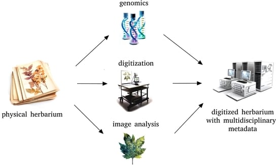
<p>Herbarium specimens are sources of a large amount of data not only related to taxonomy but also to plant phenology and biology.</p>
Full article ">Figure 2
<p>Different imaging techniques can be used for the study of herbarium collections (without damaging specimens) to collect data that can be integrated with morphological and genetic data.</p>
Full article ">
35 pages, 593 KiB  
Review
Domestic Animal Models of Central Nervous System Tumors: Focus on Meningiomas
by Michele Tomanelli, Tullio Florio, Gabriela Coronel Vargas, Aldo Pagano and Paola Modesto
Life 2023, 13(12), 2284; https://doi.org/10.3390/life13122284 - 30 Nov 2023
Cited by 2 | Viewed by 2338
Abstract
Intracranial primary tumors (IPTs) are aggressive forms of malignancies that cause high mortality in both humans and domestic animals. Meningiomas are frequent adult IPTs in humans, dogs, and cats, and both benign and malignant forms cause a decrease in life quality and survival. [...] Read more.
Intracranial primary tumors (IPTs) are aggressive forms of malignancies that cause high mortality in both humans and domestic animals. Meningiomas are frequent adult IPTs in humans, dogs, and cats, and both benign and malignant forms cause a decrease in life quality and survival. Surgery is the primary therapeutic approach to treat meningiomas, but, in many cases, it is not resolutive. The chemotherapy and targeted therapy used to treat meningiomas also display low efficacy and many side effects. Therefore, it is essential to find novel pharmacological approaches to increase the spectrum of therapeutic options for meningiomas. This review analyzes the similarities between human and domestic animal (dogs and cats) meningiomas by evaluating the molecular and histological characteristics, diagnosis criteria, and treatment options and highlighting possible research areas to identify novel targets and pharmacological approaches, which are useful for the diagnosis and therapy of this neoplasia to be used in human and veterinary medicine. Full article
18 pages, 1312 KiB  
Review
Review of Predicting Synergistic Drug Combinations
by Yichen Pan, Haotian Ren, Liang Lan, Yixue Li and Tao Huang
Life 2023, 13(9), 1878; https://doi.org/10.3390/life13091878 - 7 Sep 2023
Cited by 7 | Viewed by 5190
Abstract
The prediction of drug combinations is of great clinical significance. In many diseases, such as high blood pressure, diabetes, and stomach ulcers, the simultaneous use of two or more drugs has shown clear efficacy. It has greatly reduced the progression of drug resistance. [...] Read more.
The prediction of drug combinations is of great clinical significance. In many diseases, such as high blood pressure, diabetes, and stomach ulcers, the simultaneous use of two or more drugs has shown clear efficacy. It has greatly reduced the progression of drug resistance. This review presents the latest applications of methods for predicting the effects of drug combinations and the bioactivity databases commonly used in drug combination prediction. These studies have played a significant role in developing precision therapy. We first describe the concept of synergy. we study various publicly available databases for drug combination prediction tasks. Next, we introduce five algorithms applied to drug combinatorial prediction, which include traditional machine learning methods, deep learning methods, mathematical methods, systems biology methods and search algorithms. In the end, we sum up the difficulties encountered in prediction models. Full article
Show Figures

Figure 1

Figure 1
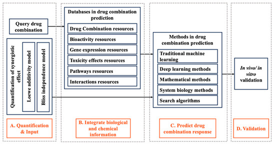
<p>The workflow of the synergy scoring system of the drug combination. In predicting drug combination response, the model calculates synergistic scores according to the quantification of synergistic effect, input biological data and chemical information using a specific synergistic quantification method. Finally, in vitro/in vivo experiments were carried out.</p>
Full article ">Figure 2
<p>The overall framework of drug combination response prediction model. The principle of predicting drug combination response is to use the information provided by the database and advanced computational models to predict the synergy score of a specific cell line for a specific drug combination.</p>
Full article ">Figure 3
<p>The isobolographic of Loewe additivity model. It shows the biological effects of drug combinations at different doses. The yellow curve shows the synergistic effect, and the green curve shows the antagonistic effect.</p>
Full article ">
25 pages, 1591 KiB  
Review
Regulatory miRNAs and lncRNAs in Skin Cancer: A Narrative Review
by Nicole Natarelli, Aleena Boby, Shaliz Aflatooni, Jasmine Thuy Tran, Michael Joseph Diaz, Kamil Taneja and Mahtab Forouzandeh
Life 2023, 13(8), 1696; https://doi.org/10.3390/life13081696 - 6 Aug 2023
Cited by 7 | Viewed by 2050
Abstract
Non-coding RNAs (ncRNAs) have a significant regulatory role in the pathogenesis of skin cancer, despite the fact that protein-coding genes have generally been the focus of research efforts in the field. We comment on the actions of long non-coding RNAs (lncRNAs) and microRNAs [...] Read more.
Non-coding RNAs (ncRNAs) have a significant regulatory role in the pathogenesis of skin cancer, despite the fact that protein-coding genes have generally been the focus of research efforts in the field. We comment on the actions of long non-coding RNAs (lncRNAs) and microRNAs (miRNAs) in the current review with an eye toward potential therapeutic treatments. LncRNAs are remarkably adaptable, acting as scaffolding, guides, or decoys to modify key signaling pathways (i.e., the Wnt/β-catenin pathway) and gene expression. As post-transcriptional gatekeepers, miRNAs control gene expression by attaching to messenger RNAs and causing their degradation or suppression during translation. Cell cycle regulation, cellular differentiation, and immunological responses are all affected by the dysregulation of miRNAs observed in skin cancer. NcRNAs also show promise as diagnostic biomarkers and prognostic indicators. Unraveling the complexity of the regulatory networks governed by ncRNAs in skin cancer offers unprecedented opportunities for groundbreaking targeted therapies, revolutionizing the landscape of dermatologic care. Full article
Show Figures

Figure 1

Figure 1
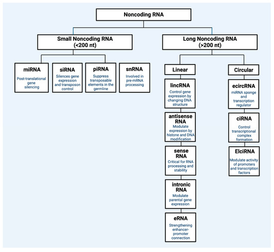
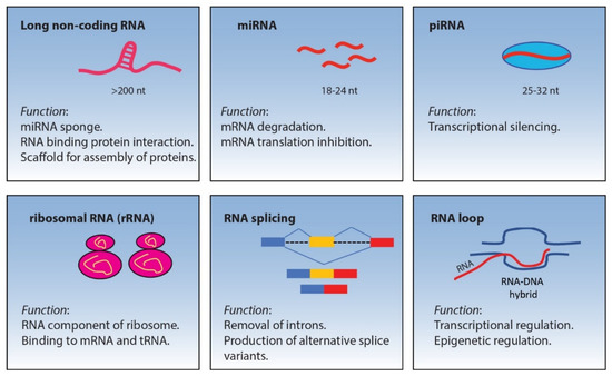
<p>Classification of main regulatory non-coding RNA types. Regulatory ncRNAs are broadly divided into two general types: small non-coding RNA (&lt;200 nucleotide) and long non-coding RNA (&gt;200 nucleotides). Within each general type, there are different subtypes of non-coding RNA classes that each have specific mechanisms of gene control. miRNA: microRNA; siRNA: small interfering RNA; pirRNA: Piwi-interacting RNA; snRNA: small nuclear RNA; lincRNA: long intergenic non-coding RNA; eRNA: enhancer RNA; ciRNA: intronic RNA; ecircRNA: exonic circular RNA; ElciRNA: exon–intron circular RNA; Nt: nucleotide. Figure created with Biorender.com.</p>
Full article ">Figure 2
<p>Pro-tumor roles of non-coding RNA in melanoma. (<b>A</b>) Tumor proliferation. (<b>B</b>) Tumor metastasis. (<b>C</b>) Tumor survival via anti-apoptotic signaling. Figure created with Biorender.com.</p>
Full article ">Figure 3
<p>Illustration of miRNA sponge therapy and antisense oligonucleotides (ASO) therapy. miRNA sponges (often circular RNAs) can be administered as nanoparticles to patients, and they contain “sticky” regions that are designed to specifically bind to target RNAs. This is a potent method of silencing RNAs. ASOs are single strand DNA segments that are specifically designed to bind RNA. These DNA-RNA complexes can be recognized by the RNase H1 complex, which in turn will degrade the RNA. Figure created with Biorender.com.</p>
Full article ">
14 pages, 839 KiB  
Review
Trends on Human Norovirus Virus-like Particles (HuNoV-VLPs) and Strategies for the Construction of Infectious Viral Clones toward In Vitro Replication
by Emilly Sion, Sharaniza Ab-Rahim and Mudiana Muhamad
Life 2023, 13(7), 1447; https://doi.org/10.3390/life13071447 - 26 Jun 2023
Cited by 1 | Viewed by 2379
Abstract
Most acute gastroenteritis (AGE) outbreaks and sporadic cases in developing countries are attributable to infection by human norovirus (HuNoV), the enteric virus mainly transmitted via fecal-contaminated water. However, it has been challenging to study HuNoV due to the lack of suitable systems to [...] Read more.
Most acute gastroenteritis (AGE) outbreaks and sporadic cases in developing countries are attributable to infection by human norovirus (HuNoV), the enteric virus mainly transmitted via fecal-contaminated water. However, it has been challenging to study HuNoV due to the lack of suitable systems to cultivate and replicate the virus, hindering the development of treatments and vaccines. Researchers have been using virus-like particles (VLPs) and infectious viral clones to overcome this challenge as alternatives to fresh virus isolates in various in vitro and ex vivo models. VLPs are multiprotein structures that mimic the wild-type virus but cannot replicate in host cells due to the lack of genetic materials for replication, limiting downstream analysis of the virus life cycle and pathogenesis. The development of in vitro cloning systems has shown promise for HuNoV replication studies. This review discusses the approaches for constructing HuNoV-VLPs and infectious viral clones, the techniques involved, and the challenges faced. It also highlights the relationship between viral genes and their protein products and provides a perspective on technical considerations for producing efficient HuNoV-VLPs and infectious viral clones, which could substitute for native human noroviruses in future studies. Full article
Show Figures

Figure 1

Figure 1
<p>Genome organization of human norovirus (HuNoV). The ORF1 expresses nonstructural proteins starting from 5′ end: p48, NTPase, p22, VPg, protease, and RdRp. The ORF2 encodes for the major structural protein VP1, while ORF3 encodes for the minor structural protein VP2. A subgenomic expressing VP1 and VP2 is linked to VPg as shown below the genomic RNA with the link illustrated as the round shape beside VP1. Adapted from [<a href="#B18-life-13-01447" class="html-bibr">18</a>].</p>
Full article ">Figure 2
<p>Overview of the common cloning methods. (<b>A</b>) The plasmid vector is linearized by digestion with restriction enzyme (RE) on multiple cloning sites producing “sticky ends”. Foreign DNA molecules or inserts are digested with the same RE producing “sticky ends” complimentary to the plasmid vector. The transformed vector is cloned into competent cells for selection of cells with transformed vectors that can grow on the selective media. The illustration was adapted from [<a href="#B65-life-13-01447" class="html-bibr">65</a>]. (<b>B</b>) The DNA insert with 3′ tail consists of a 3′-OH generated by PCR. Blunt-end linearized vector is prepared by ddT tailing with terminal transferase compatible with the insert ends. Ligation of the ddT-tailed vector and DNA insert are due to the formation of phosphodiester bonds between the 5′-P of the vector and the 3′-OH group of the PCR product. Successfully ligated product can be transformed into competent cells. Growth colony on selective media indicates cells carrying the transformed vector. The TA cloning illustration was adapted from [<a href="#B66-life-13-01447" class="html-bibr">66</a>]. (<b>C</b>) Linearized plasmid vector and DNA inserts with respective overlapping regions are assembled in a single isothermal reaction. This method requires a 20–80 bp sequence overlap at the end of the DNA elements to be assembled. The assembled product can be stored in −20 °C or directly transformed into competent bacterial cells. Illustration was adapted from Gibson Assembly in New England Biolabs, n.d. Retrieved 16 December 2021 from <a href="https://international.neb.com/applications/cloning-and-synthetic-biology/dna-assembly-and-cloning/gibson-assembly" target="_blank">https://international.neb.com/applications/cloning-and-synthetic-biology/dna-assembly-and-cloning/gibson-assembly</a>. Copyright 2021 from New England Biolabs.</p>
Full article ">
13 pages, 2407 KiB  
Review
Research Trends in C-Terminal Domain Nuclear Envelope Phosphatase 1
by Harikrishna Reddy Rallabandi, Haewon Choi, Hyunseung Cha and Young Jun Kim
Life 2023, 13(6), 1338; https://doi.org/10.3390/life13061338 - 7 Jun 2023
Cited by 4 | Viewed by 1804
Abstract
C-terminal domain nuclear envelope phosphatase 1 (CTDNEP1, formerly Dullard) is a member of the newly emerging protein phosphatases and has been recognized in neuronal cell tissues in amphibians. It contains the phosphatase domain in the C-terminal, and the sequences are conserved in various [...] Read more.
C-terminal domain nuclear envelope phosphatase 1 (CTDNEP1, formerly Dullard) is a member of the newly emerging protein phosphatases and has been recognized in neuronal cell tissues in amphibians. It contains the phosphatase domain in the C-terminal, and the sequences are conserved in various taxa of organisms. CTDNEP1 has several roles in novel biological activities such as neural tube development in embryos, nuclear membrane biogenesis, regulation of bone morphogenetic protein signaling, and suppression of aggressive medulloblastoma. The three-dimensional structure of CTDNEP1 and the detailed action mechanisms of CTDNEP1’s functions have yet to be determined for several reasons. Therefore, CTDNEP1 is a protein phosphatase of interest due to recent exciting and essential works. In this short review, we summarize the presented biological roles, possible substrates, interacting proteins, and research prospects of CTDNEP1. Full article
Show Figures

Figure 1

Figure 1
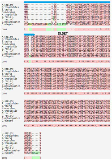
<p>The multiple sequence alignments of CTDNEP1 homologs produced using T-Coffee (<a href="https://tcoffee.crg.eu/" target="_blank">https://tcoffee.crg.eu/</a>, accessed on 19 March 2023). GenBank accession numbers corresponding to the homologs are given below; Homo sapiens, NP_0001137247.1; Pan troglodytes, XP_511976.3; Bos taurus, NP_001039491.1; Mus musculus, NP_080293.1; Xenopus tropicalis, NP_001017177.1; Danio rerio, NP_001007310.1, NP_001007441.1; Drosophila melanogaster, NP_608449.1; Caenorhabditis elegans, NP_001254123.1. DLDET represents the active site sequences, and the transmembrane region is presented as a blue bar. The dark pink colors are reliable alignments, while the green colors are unreliable alignments. The yellow colors are moderately reliable alignments. The cons means the degree of conservation observed in each column, which is denoted by the following symbols; an * (asterisk) indicates positions which have a single, fully conserved residue; a : (colon) indicates conservation between groups of strongly similar properties; a . (period) indicates conservation between groups of weakly similar properties.</p>
Full article ">Figure 2
<p>The structural comparison of human CTDNEP1 (<b>d</b>), a model structure predicted using alphafold2) with human CTDSP1 (<b>a</b>), pdb ID = 2ghq, a complex structure of the green-colored CTD phosphatase domain of CTDSP1 with the other colored CTD peptide of RNAPII), human CTDSP2 (<b>b</b>), pdb ID = 2q5e, an apo structure of the green-colored CTD phosphatase domain of CTDSP2), and human CTDSPL (<b>c</b>), pdb ID = 2hhl, a complex structure of the green-colored CTD phosphatase domain of CTDSPL with the other colored phosphatase inhibitor). In the model structure of CTDNEP1, the blue color represents very high per-residue confidence (pLDDT &gt; 90), and the sky-blue color represents high confidence (90 &gt; pLDDT &gt; 70). The yellow color represents low confidence (70 &gt; pLDDT &gt; 50).</p>
Full article ">
13 pages, 2153 KiB  
Review
Tensins in Kidney Function and Diseases
by Chien-Wei Huang and Su Hao Lo
Life 2023, 13(6), 1244; https://doi.org/10.3390/life13061244 - 24 May 2023
Cited by 1 | Viewed by 1848
Abstract
Tensins are focal adhesion proteins that regulate various biological processes, such as mechanical sensing, cell adhesion, migration, invasion, and proliferation, through their multiple binding activities that transduce critical signals across the plasma membrane. When these molecular interactions and/or mediated signaling are disrupted, cellular [...] Read more.
Tensins are focal adhesion proteins that regulate various biological processes, such as mechanical sensing, cell adhesion, migration, invasion, and proliferation, through their multiple binding activities that transduce critical signals across the plasma membrane. When these molecular interactions and/or mediated signaling are disrupted, cellular activities and tissue functions are compromised, leading to disease development. Here, we focus on the significance of the tensin family in renal function and diseases. The expression pattern of each tensin in the kidney, their roles in chronic kidney diseases, renal cell carcinoma, and their potentials as prognostic markers and/or therapeutic targets are discussed in this review. Full article
Show Figures

Figure 1

Figure 1
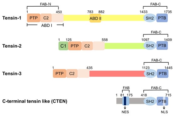
<p>Domain structures of human tensins. Tensin-1 (TNS1), tensin-2 (TNS2), and tensin-3 (TNS3) have similar domain structures, including the PTP, C2, SH2, PTB, ABD, and FAB domains. There are two independent FAB regions: FAB-N domains contain the PTP and C2 domains at the N-terminal region and FAB-C overlaps with the SH2 and PTB domains at the C-terminus. ABD I, which interacts directly with actin filaments, is located at the N-terminus and overlaps with FAB-N. ABD II binds to the barbed end of the actin filaments. The protein kinase C conserved region 1 domain (C1 domain) is found in TNS2 with an uncertain function. CTEN harbors the SH2-PTB tandem domain and contains the FAB-C domain. Although CTEN also features a second FAB domain in its N-terminal region, its amino acid sequence is different from those of FAB-N in other tensins [<a href="#B14-life-13-01244" class="html-bibr">14</a>]. Moreover, there is a nuclear export sequence (NES) localized within this unique FAB site of CTEN and a nuclear localization sequence (NLS) within CTEN’s PTB domain [<a href="#B14-life-13-01244" class="html-bibr">14</a>].</p>
Full article ">Figure 2
<p><span class="html-italic">Tns1</span>-KO mice develop features of cystic kidney disease. <span class="html-italic">Tns1</span>-KO kidneys isolated from 3-month-old (<b>A</b>,<b>B</b>) or 10-month-old (<b>C</b>,<b>D</b>) mice were processed for H&amp;E staining (<b>A</b>,<b>B</b>), Sirius Red staining (<b>D</b>), or general morphology (<b>C</b>), showing dilated tubules (black arrows), interstitial infiltration (arrowhead), fibrosis (blue arrows), and cystic kidneys. Representative images of <span class="html-italic">Tns1</span> WT (<b>E</b>) or KO (<b>F</b>) MDCK in 3D Matrigel culture for 5 days and stained for actin.</p>
Full article ">Figure 3
<p><span class="html-italic">Tns2</span>-KO FVB mice develop features of glomerulonephritis. Kidneys isolated from 9-week-old (<b>A</b>) or 6-week-old (<b>B</b>,<b>C</b>) <span class="html-italic">Tns2</span>-KO FBV mice, generated in the laboratory using <span class="html-italic">Tns2</span>-KO embryonic stem cells from the Knockout Mouse Programme (<a href="http://www.mousephenotype.org" target="_blank">www.mousephenotype.org</a>, accessed on 10 October 2015), were processed and stained with H&amp;E, showing mesangial proliferation (black arrows), segmental glomerulosclerosis (arrowhead), tubular dilatation (asterisk), cast (blue dot), and interstitial infiltration (red arrow). These defects are mouse-strain-dependent and are not developed in the kidneys of <span class="html-italic">Tns2</span>-KO C57BL6 mice, even at 2 years of age (<b>D</b>).</p>
Full article ">Figure 4
<p>Cumulative survival rate of patients with RCC by tensin status. The Kaplan–Meier curves reveal the cumulative survival rate among patients with CCC (N = 530) and those with PCC (N = 288), stratified according to the status of each tensin. The <span class="html-italic">p</span> values are the result of the log-rank test for the comparison between the two groups. High <span class="html-italic">CTEN</span> (HR = 1.63, <span class="html-italic">p</span> = 0.0023), low <span class="html-italic">TNS1</span> (HR = 0.38, <span class="html-italic">p</span> &lt; 0.001), and low <span class="html-italic">TNS3</span> (HR = 0.45, <span class="html-italic">p</span> &lt; 0.001) increase the risk of mortality significantly in patients with CCC. High <span class="html-italic">TNS2</span> is significantly associated with poor survival in those with PCC (HR = 2, <span class="html-italic">p</span> = 0.026). The association between <span class="html-italic">TNS1</span>, <span class="html-italic">TNS3</span>, and <span class="html-italic">TNS4</span> and survival in patients with PCC is not significant.</p>
Full article ">
36 pages, 1080 KiB  
Review
A Comprehensive Review on Weight Loss Associated with Anti-Diabetic Medications
by Fatma Haddad, Ghadeer Dokmak, Maryam Bader and Rafik Karaman
Life 2023, 13(4), 1012; https://doi.org/10.3390/life13041012 - 14 Apr 2023
Cited by 18 | Viewed by 15109
Abstract
Obesity is a complex metabolic condition that can have a negative impact on one’s health and even result in mortality. The management of obesity has been addressed in a number of ways, including lifestyle changes, medication using appetite suppressants and thermogenics, and bariatric [...] Read more.
Obesity is a complex metabolic condition that can have a negative impact on one’s health and even result in mortality. The management of obesity has been addressed in a number of ways, including lifestyle changes, medication using appetite suppressants and thermogenics, and bariatric surgery for individuals who are severely obese. Liraglutide and semaglutide are two of the five Food and Drug Administration (FDA)-approved anti-obesity drugs that are FDA-approved agents for the treatment of type 2 diabetes mellitus (T2DM) patients. In order to highlight the positive effects of these drugs as anti-obesity treatments, we analyzed the weight loss effects of T2DM agents that have demonstrated weight loss effects in this study by evaluating clinical studies that were published for each agent. Many clinical studies have revealed that some antihyperglycemic medications can help people lose weight, while others either cause weight gain or neutral results. Acarbose has mild weight loss effects and metformin and sodium-dependent glucose cotransporter proteins-2 (SGLT-2) inhibitors have modest weight loss effects; however, some glucagon-like peptide-1 (GLP-1) receptor agonists had the greatest impact on weight loss. Dipeptidyl peptidase 4 (DPP-4) inhibitors showed a neutral or mild weight loss effect. To sum up, some of the GLP-1 agonist drugs show promise as weight-loss treatments. Full article
Show Figures

Figure 1

Figure 1
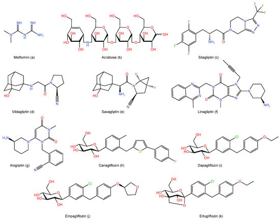
<p>The chemical structures of: (<b>a</b>) Metformin, (<b>b</b>) Acarbose, (<b>c</b>) Sitagliptin, (<b>d</b>) Vildagliptin, (<b>e</b>) Saxagliptin, (<b>f</b>) Linagliptin, (<b>g</b>) Alogliptin, (<b>h</b>) Canagliflozin, (<b>i</b>) Dapagliflozin, (<b>j</b>) Empagliflozin, and (<b>k</b>) Ertugliflozin.</p>
Full article ">Figure 2
<p>The primary mechanism of action of the biguanide, alpha-glucosidase inhibitors, dipeptidyl peptidase-4 (DPP-4) inhibitors, sodium-dependent glucose cotransporter proteins-2 (SGLT-2) inhibitors, and glucagon-like peptide-1 (GLP-1) agonists.</p>
Full article ">
18 pages, 2024 KiB  
Review
Current Views on Infective Endocarditis: Changing Epidemiology, Improving Diagnostic Tools and Centering the Patient for Up-to-Date Management
by Giovanni Cimmino, Roberta Bottino, Tiziana Formisano, Massimiliano Orlandi, Daniele Molinari, Simona Sperlongano, Pasquale Castaldo, Saverio D’Elia, Andreina Carbone, Alberto Palladino, Lavinia Forte, Francesco Coppolino, Michele Torella and Nicola Coppola
Life 2023, 13(2), 377; https://doi.org/10.3390/life13020377 - 30 Jan 2023
Cited by 18 | Viewed by 9236
Abstract
Infective endocarditis (IE) is a rare but potentially life-threatening disease, sometimes with longstanding sequels among surviving patients. The population at high risk of IE is represented by patients with underlying structural heart disease and/or intravascular prosthetic material. Taking into account the increasing number [...] Read more.
Infective endocarditis (IE) is a rare but potentially life-threatening disease, sometimes with longstanding sequels among surviving patients. The population at high risk of IE is represented by patients with underlying structural heart disease and/or intravascular prosthetic material. Taking into account the increasing number of intravascular and intracardiac procedures associated with device implantation, the number of patients at risk is growing too. If bacteremia develops, infected vegetation on the native/prosthetic valve or any intracardiac/intravascular device may occur as the final result of invading microorganisms/host immune system interaction. In the case of IE suspicion, all efforts must be focused on the diagnosis as IE can spread to almost any organ in the body. Unfortunately, the diagnosis of IE might be difficult and require a combination of clinical examination, microbiological assessment and echocardiographic evaluation. There is a need of novel microbiological and imaging techniques, especially in cases of blood culture-negative. In the last few years, the management of IE has changed. A multidisciplinary care team, including experts in infectious diseases, cardiology and cardiac surgery, namely, the Endocarditis Team, is highly recommended by the current guidelines. Full article
Show Figures

Figure 1

Figure 1
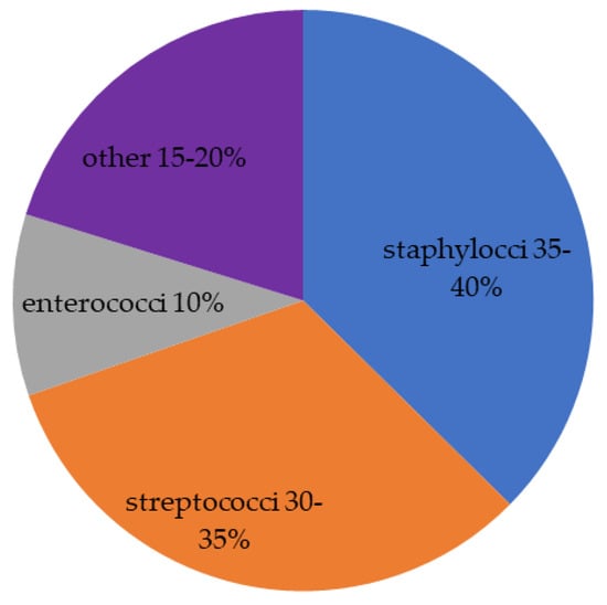
<p>Distribution of pathogens mainly involved in IE. The last group includes HACEK group (2%), fungi, polymicrobial infection and, rarely, aerobic Gram-negative bacilli.</p>
Full article ">Figure 2
<p>Distribution of pathogens in different clinical settings. (<b>A</b>) cardiac device related-infective endocarditis; the other group includes <span class="html-italic">Candida</span> spp., other fungi, Gram-negative bacilli and polymicrobial. (<b>B</b>) transcatheter aortic valve implantation (TAVI); the other group includes Gram-negative bacille, Moraxella, candida albicans, Histoplasma and corynebacterium. (<b>C</b>) right-sided infective endocarditis; the other group includes streptococci, coagulase-negative staphylococcal, pseudomonas aeruginosa and fungi. (<b>D</b>) immunosuppressive therapy in solid organ transplantation; the other group includes Gram-negative bacilli, Corynebacteria, Clostridium ramosum, Pseudallescheria boydii, Nocardia asteroids and Polymicrobial.</p>
Full article ">Figure 3
<p>IE of both tricuspid and mitral valve. Vegetations are well-imaged by TTE, which is the first imaging tool in diagnosing IE (<b>A</b>). Their presence, size and location are confirmed by TOE (<b>B</b>,<b>C</b>,<b>E</b>). Both vegetations are associated with significant valve regurgitations (<b>D</b>,<b>F</b>). IE: infective endocarditis; TTE: transthoracic echocardiography; TOE: transesophageal echocardiography.</p>
Full article ">Figure 4
<p>Diagnostic imaging tools in suspected IE. Structural complications include abscess, pseudoaneurysm, intracardiac fistula, valvular perforation or aneurysm and new dehiscence of prosthetic valve. CT: computed tomography; FDG: fluorodeoxyglucose; IE: infective endocarditis; PET: positron emission tomography; SPECT: single photon emission computed tomography; TOE: transesophageal echocardiography; TTE: transthoracic echocardiography.</p>
Full article ">Figure 5
<p>Schematic view of Endocarditis Team. The continuous line indicates the main specialists involved in the decision-making process where the patient is in the center. Discontinuous line indicates the other medical specialists that might be involved according to the single case.</p>
Full article ">
22 pages, 691 KiB  
Review
A Synopsis of Current Theories on Drug-Induced Nephrotoxicity
by Lukasz Dobrek
Life 2023, 13(2), 325; https://doi.org/10.3390/life13020325 - 24 Jan 2023
Cited by 12 | Viewed by 8881
Abstract
The overriding goal of the treatment of patients is its effectiveness and safety. However, all medications currently being used also exert some adverse pharmaceutical reactions, which may be regarded as an unintended but inevitable cost of pharmacotherapy. The kidney, as the main organ [...] Read more.
The overriding goal of the treatment of patients is its effectiveness and safety. However, all medications currently being used also exert some adverse pharmaceutical reactions, which may be regarded as an unintended but inevitable cost of pharmacotherapy. The kidney, as the main organ that eliminates xenobiotics, is an organ especially predisposed and vulnerable to the toxic effects of drugs and their metabolites during their excretion from the body. Moreover, some drugs (e.g., aminoglycosides, cyclosporin A, cisplatin, amphotericin B, and others) have a “preferential” nephrotoxicity potential, and their use is associated with an increased risk of kidney damage. Drug nephrotoxicity is, therefore, both a significant problem and a complication of pharmacotherapy. It should be noted that, currently, there is no generally recognized definition of drug-induced nephrotoxicity and no clear criteria for its diagnosis. This review briefly describes the epidemiology and diagnosis of drug-induced nephrotoxicity and characterizes its pathomechanisms, including immunological and inflammatory disturbances, altered kidney blood flow, tubulointerstitial injury, increased lithogenesis–crystal nephropathy, rhabdomyolysis, and thrombotic microangiopathy. The study also lists the basic drugs with nephrotoxicity potential and provides a short overview of the preventive methods for reducing the risk of drug-related kidney damage developing. Full article
Show Figures

Figure 1

Figure 1
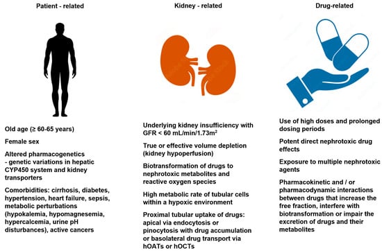
<p>Risk factors for drug-induced kidney disease development [<a href="#B8-life-13-00325" class="html-bibr">8</a>,<a href="#B9-life-13-00325" class="html-bibr">9</a>,<a href="#B10-life-13-00325" class="html-bibr">10</a>]. hOAT—human organic anion transporters; hOCTs—human organic cation transporters.</p>
Full article ">

2022

Jump to: 2024, 2023, 2021

10 pages, 230 KiB  
Review
Infectious Risks Related to Umbilical Venous Catheter Dwell Time and Its Replacement in Newborns: A Narrative Review of Current Evidence
by Lucia Corso, Martina Buttera, Francesco Candia, Francesca Sforza, Katia Rossi, Licia Lugli, Francesca Miselli, Luca Bedetti, Cecilia Baraldi, Laura Lucaccioni, Lorenzo Iughetti and Alberto Berardi
Life 2023, 13(1), 123; https://doi.org/10.3390/life13010123 - 31 Dec 2022
Cited by 10 | Viewed by 3491
Abstract
The use of umbilical venous catheters (UVCs) has become the standard of care in the neonatal intensive care unit (NICU) to administer fluids, medications and parenteral nutrition. However, it is well known that UVCs can lead to some serious complications, both mechanical and [...] Read more.
The use of umbilical venous catheters (UVCs) has become the standard of care in the neonatal intensive care unit (NICU) to administer fluids, medications and parenteral nutrition. However, it is well known that UVCs can lead to some serious complications, both mechanical and infective, including CLABSI (Central Line-Associated Bloodstream Infections). Most authors recommend removing UVC within a maximum of 14 days from its placement. However, the last Infusion Therapy Standards of Practice (INS) guidelines recommends limiting the UVC dwell time to 7 to 10 days, to reduce risks of infectious and thrombotic complications. These guidelines also suggest as an infection prevention strategy to remove UVC after 4 days, followed by the insertion of a PICC if a central line is still needed. Nevertheless, the maximum UVC dwell time to reduce the risk of CLABSI is still controversial, as well as the time of its replacement with a PICC. In this study we reviewed a total of 177 articles, found by using the PubMed database with the following search strings: “UVC AND neonates”, “(neonate* OR newborn*) AND (UVC OR central catheter*) AND (infection*)”. We also analyze the INS guidelines to provide the reader an updated overview on this topic. The purpose of this review is to give updated information on CVCs infectious risks by examining the literature in this field. These data could help clinicians in deciding the best time to remove or to replace the UVC with a PICC, to reduce CLABSIs risk. Despite the lack of strong evidence, the risk of CLABSI seems to be minimized when UVC is removed/replaced within 7 days from insertion and this indication is emerging from more recent and larger studies. Full article
22 pages, 3371 KiB  
Review
Application of Nanoparticles: Diagnosis, Therapeutics, and Delivery of Insulin/Anti-Diabetic Drugs to Enhance the Therapeutic Efficacy of Diabetes Mellitus
by Tilahun Ayane Debele and Yoonjee Park
Life 2022, 12(12), 2078; https://doi.org/10.3390/life12122078 - 11 Dec 2022
Cited by 9 | Viewed by 5219
Abstract
Diabetes mellitus (DM) is a chronic metabolic disorder of carbohydrates, lipids, and proteins due to a deficiency of insulin secretion or failure to respond to insulin secreted from pancreatic cells, which leads to high blood glucose levels. DM is one of the top [...] Read more.
Diabetes mellitus (DM) is a chronic metabolic disorder of carbohydrates, lipids, and proteins due to a deficiency of insulin secretion or failure to respond to insulin secreted from pancreatic cells, which leads to high blood glucose levels. DM is one of the top four noncommunicable diseases and causes of death worldwide. Even though great achievements were made in the management and treatment of DM, there are still certain limitations, mainly related to the early diagnosis, and lack of appropriate delivery of insulin and other anti-diabetic agents. Nanotechnology is an emerging field in the area of nanomedicine and NP based anti-diabetic agent delivery is reported to enhance efficacy by increasing bioavailability and target site accumulation. Moreover, theranostic NPs can be used as diagnostic tools for the early detection and prevention of diseases owing to their unique biological, physiochemical, and magnetic properties. NPs have been synthesized from a variety of organic and inorganic materials including polysaccharides, dendrimers, proteins, lipids, DNA, carbon nanotubes, quantum dots, and mesoporous materials within the nanoscale size. This review focuses on the role of NPs, derived from organic and inorganic materials, in the diagnosis and treatment of DM. Full article
Show Figures

Figure 1

Figure 1
<p>The etiology, pathophysiology, and conventional treatment methods of DM.</p>
Full article ">Figure 2
<p>Classification, biomedical applications, and advantages of nanoparticles (NPs).</p>
Full article ">Figure 3
<p>(<b>a</b>) Scheme of insulin encapsulation in polymersomes, (<b>b</b>) Insulin release profile at pH 7.4 from DEX5000-PLGA13000 polymersomes, (<b>c</b>) Insulin release profile at pH 1.2 from DEX5000-PLGA13000 polymersomes, (<b>d</b>) blood glucose level and (<b>e</b>) blood insulin concentration of diabetic rats after subcutaneous insulin solution injection at a dose of 5 IU/kg/b.w., orally administrated insulin solution at a dose of 100 IU/kg/b.w and orally administrated F7 formulation at a dose of 100 IU/kg/b.w. Reproduced with permission from Elsevier [<a href="#B89-life-12-02078" class="html-bibr">89</a>].</p>
Full article ">Figure 4
<p>(<b>a</b>) Synthesis of chitosan-derivative, (<b>b</b>) Schematic illustration of chitosan-based multifunctional nanocarriers (<b>c</b>) The insulin release in vitro, from nanocarriers against SGF (pH 1.2) and SIF (pH 6.8) at different concentrations of glucose (at T = 37 °C) (<b>d</b>) Serum insulin level in diabetic rats following oral administration of free insulin, insulin solution and insulin-loaded nanocarriers (<b>e</b>) Blood glucose levels in diabetic rats following oral administration of saline, insulin-loaded nanocarriers, free insulin, and insulin solution. Reproduced with permission from Elsevier [<a href="#B98-life-12-02078" class="html-bibr">98</a>].</p>
Full article ">Figure 5
<p>(<b>a</b>) Release kinetics from rapid-release (high acyclic acetal content) and extended-release (high cyclic acetal content) Ac-dex NPs, (<b>b</b>) Rate of degradation of Ac-dex NPs by percent cyclic modification, as determined by the concentration of soluble dextran in the supernatant. (<b>c</b>) Schematic of glucose-responsive insulin release from acid-degradable Ac-dex NPs, (<b>d</b>) Average relative diameter of C55NPs in acetate buffer showing a reduction in size over time. Inset: cryo-transmission electron micrograph of NPs at time 0. (<b>e</b>) Insulin release from C55NPs or C71NPs incubated in either acetate buffer (pH 4.7) or phosphate-buffered saline (PBS, pH 7.4) at 37 °C, (<b>f</b>) Blood glucose levels of streptozotocin-induced type 1 diabetic mice following administration of empty NPs, 3 IU/kg naked insulin, or 14.4 IU/kg insulin in Ac-dex NPs. Arrows represent intraperitoneal glucose tolerance tests (GTTs, 1.5 g/kg) every 3 h. Statistical significance is indicated by **** <span class="html-italic">p</span> &lt; 0.0001. Reproduced with permission from the American Chemical Society [<a href="#B105-life-12-02078" class="html-bibr">105</a>].</p>
Full article ">Figure 6
<p>(<b>a</b>) Schematic illustration for the complexation and glucose-responsive disintegration of the PEG-b-P(AA-co-APBA)/P(AA-co-AGA) complex micelles. Glucose-responsiveness of the complex micelles in the presence of glucose with different concentrations, (<b>b</b>) 2, (<b>c</b>) 5, (<b>d</b>) 20, and (<b>e</b>) 50 g/L in aqueous solutions of PBS 7.4 in terms of normalized light scattering intensity as a function of time. Reproduced with permission from the American Chemical Society [<a href="#B109-life-12-02078" class="html-bibr">109</a>].</p>
Full article ">Figure 7
<p>In vivo study of gliclazide loaded PLGA SGNCs formulation, (<b>a</b>) Pharmacodynamic responses of pure gliclazide, placebo and optimized formulation, SGNCF1 on type II diabetic rats, (<b>b</b>) Plasma mean gliclazide concentration–time curves after a single oral dosing of pure gliclazide and optimized formulation, SGNCF1 on type 2 diabetic rats, (<b>c</b>) Pharmacokinetic–pharmacodynamic correlation plot of gliclazide loaded PLGA SGNC formulation on type 2 diabetic rats. Reproduced with permission from Springer nature [<a href="#B115-life-12-02078" class="html-bibr">115</a>].</p>
Full article ">
12 pages, 263 KiB  
Review
Quantitative Assessment of Asbestos Fibers in Normal and Pathological Peritoneal Tissue—A Scoping Review
by Yohama Caraballo-Arias, Carlotta Zunarelli, Paola Caffaro, Francesco Roccuzzo, Mattia Roberto Nocilla, Maria Chiara Imperiale, Clara Romano, Paolo Boffetta and Francesco Saverio Violante
Life 2022, 12(12), 1969; https://doi.org/10.3390/life12121969 - 24 Nov 2022
Cited by 2 | Viewed by 1857
Abstract
Peritoneal tissue is the second most affected site by malignant mesothelioma linked to asbestos exposure. This scoping review aims to summarize the findings of the studies in which asbestos fibers in the peritoneum were quantified by electron microscopy, occasionally associated with spectroscopy, both [...] Read more.
Peritoneal tissue is the second most affected site by malignant mesothelioma linked to asbestos exposure. This scoping review aims to summarize the findings of the studies in which asbestos fibers in the peritoneum were quantified by electron microscopy, occasionally associated with spectroscopy, both in neoplastic and non-neoplastic tissue. The 9 studies selected comprised 62 cases, out of whom 100 samples were analyzed. Asbestos fibers were detected in 58 samples (58%). In addition, 28 cases had diagnosis of peritoneal mesothelioma. For 32 cases, a lung tumor sample was available: 28/32 samples analyzed presented asbestos fibers; 18/32 reported amphiboles with a range from not detected to 14.2 million fibers per gram of dry tissue (mfgdt); 18/32 reported chrysotile, with a range of 0 to 90 mfgdt. The studies were heterogeneous for type of samples, analytical technology, and circumstances of exposure to asbestos. To evaluate asbestos fibers in the peritoneum and to better understand the association between asbestos exposure and malignant peritoneal mesothelioma, it is desirable that the search for asbestos fibers becomes a routine process every time peritoneal tissue is accessible. Full article
11 pages, 1144 KiB  
Review
Effectiveness of CPR in Hypogravity Conditions—A Systematic Review
by Remco Overbeek, Jan Schmitz, Lucas Rehnberg, Yacine Benyoucef, Fabian Dusse, Thais Russomano and Jochen Hinkelbein
Life 2022, 12(12), 1958; https://doi.org/10.3390/life12121958 - 23 Nov 2022
Cited by 2 | Viewed by 2418
Abstract
(1) Background: Cardiopulmonary resuscitation (CPR), as a form of basic life support, is critical for maintaining cardiac and cerebral perfusion during cardiac arrest, a medical condition with high expected mortality. Current guidelines emphasize the importance of rapid recognition and prompt initiation of high-quality [...] Read more.
(1) Background: Cardiopulmonary resuscitation (CPR), as a form of basic life support, is critical for maintaining cardiac and cerebral perfusion during cardiac arrest, a medical condition with high expected mortality. Current guidelines emphasize the importance of rapid recognition and prompt initiation of high-quality CPR, including appropriate cardiac compression depth and rate. As space agencies plan missions to the Moon or even to explore Mars, the duration of missions will increase and with it the chance of life-threatening conditions requiring CPR. The objective of this review was to examine the effectiveness and feasibility of chest compressions as part of CPR following current terrestrial guidelines under hypogravity conditions such as those encountered on planetary or lunar surfaces; (2) Methods: A systematic literature search was conducted by two independent reviewers (PubMed, Cochrane Register of Controlled Trials, ResearchGate, National Aeronautics and Space Administration (NASA)). Only controlled trials conducting CPR following guidelines from 2010 and after with advised compression depths of 50 mm and above were included; (3) Results: Four different publications were identified. All studies examined CPR feasibility in 0.38 G simulating the gravitational force on Mars. Two studies also simulated hypogravity on the Moon with a force of 0.17 G/0,16 G. All CPR protocols consisted of chest compressions only without ventilation. A compression rate above 100/s could be maintained in all studies and hypogravity conditions. Two studies showed a significant reduction of compression depth in 0.38 G (−7.2 mm/−8.71 mm) and 0.17 G (−12.6 mm/−9.85 mm), respectively, with nearly similar heart rates, compared to 1 G conditions. In the other two studies, participants with higher body weight could maintain a nearly adequate mean depth while effort measured by heart rate (+23/+13.85 bpm) and VO2max (+5.4 mL·kg−1·min−1) increased significantly; (4) Conclusions: Adequate CPR quality in hypogravity can only be achieved under increased physical stress to compensate for functional weight loss. Without this extra effort, the depth of compression quickly falls below the guideline level, especially for light-weight rescuers. This means faster fatigue during resuscitation and the need for more frequent changes of the resuscitator than advised in terrestrial guidelines. Alternative techniques in the straddling position should be further investigated in hypogravity. Full article
Show Figures

Figure 1

Figure 1
<p>PRISMA flow diagram, adapted under the terms of the Creative Commons Attribution License from ‘The PRISMA 2020 statement: An updated guideline for reporting systematic reviews’ [<a href="#B16-life-12-01958" class="html-bibr">16</a>], 2021, Page, M.J.</p>
Full article ">Figure 2
<p>Body-suspension device [<a href="#B17-life-12-01958" class="html-bibr">17</a>,<a href="#B18-life-12-01958" class="html-bibr">18</a>,<a href="#B19-life-12-01958" class="html-bibr">19</a>], reprinted with permission from ‘Extraterrestrial CPR and Its Applications in Terrestrial Medicine’ [<a href="#B21-life-12-01958" class="html-bibr">21</a>]. 2010, Russomano T.</p>
Full article ">Figure 3
<p>The Seated-Arm-Lock method [<a href="#B37-life-12-01958" class="html-bibr">37</a>].</p>
Full article ">
28 pages, 1657 KiB  
Review
The Innovative Informatics Approaches of High-Throughput Technologies in Livestock: Spearheading the Sustainability and Resiliency of Agrigenomics Research
by Godagama Gamaarachchige Dinesh Suminda, Mrinmoy Ghosh and Young-Ok Son
Life 2022, 12(11), 1893; https://doi.org/10.3390/life12111893 - 15 Nov 2022
Cited by 3 | Viewed by 3535
Abstract
For more than a decade, next-generation sequencing (NGS) has been emerging as the mainstay of agrigenomics research. High-throughput technologies have made it feasible to facilitate research at the scale and cost required for using this data in livestock research. Scale frameworks of sequencing [...] Read more.
For more than a decade, next-generation sequencing (NGS) has been emerging as the mainstay of agrigenomics research. High-throughput technologies have made it feasible to facilitate research at the scale and cost required for using this data in livestock research. Scale frameworks of sequencing for agricultural and livestock improvement, management, and conservation are partly attributable to innovative informatics methodologies and advancements in sequencing practices. Genome-wide sequence-based investigations are often conducted worldwide, and several databases have been created to discover the connections between worldwide scientific accomplishments. Such studies are beginning to provide revolutionary insights into a new era of genomic prediction and selection capabilities of various domesticated livestock species. In this concise review, we provide selected examples of the current state of sequencing methods, many of which are already being used in animal genomic studies, and summarize the state of the positive attributes of genome-based research for cattle (Bos taurus), sheep (Ovis aries), pigs (Sus scrofa domesticus), horses (Equus caballus), chickens (Gallus gallus domesticus), and ducks (Anas platyrhyncos). This review also emphasizes the advantageous features of sequencing technologies in monitoring and detecting infectious zoonotic diseases. In the coming years, the continued advancement of sequencing technologies in livestock agrigenomics will significantly influence the sustained momentum toward regulatory approaches that encourage innovation to ensure continued access to a safe, abundant, and affordable food supplies for future generations. Full article
Show Figures

Figure 1

Figure 1
<p>A symmetrical illustration of the period and the development of sequencing platforms.</p>
Full article ">Figure 2
<p>A comprehensive overview of the emergence of next-generation sequencing and the timeline of the valuable domestic animal genome findings.</p>
Full article ">Figure 3
<p>Mutation rates of viruses represent the rate of virus evolution. The average mutation rate of the dsDNA viruses is the lowest owing to the proofreading activity of the harbored DNA polymerase (1.38 × 10<sup>−7</sup> nucleotide substitutions/site/year in Herpes virus). The ssRNA viruses show the highest mutation rates (2.0 × 10<sup>−3</sup> nucleotide substitutions/site/year in the Ebola virus, 0.8–2.38 × 10<sup>−3</sup> in coronaviruses, and 1.21 × 10<sup>−2</sup> in norovirus). The RNA-dependent RNA and DNA polymerases are more likely to cause mutation than DNA-dependent DNA polymerases as they lack proofreading activity. The mutation rate of reverse transcriptase is higher than that of DNA polymerase; however, RNA viruses show more mutations than retroviruses.</p>
Full article ">
21 pages, 2398 KiB  
Review
M. tuberculosis Transcription Machinery: A Review on the Mycobacterial RNA Polymerase and Drug Discovery Efforts
by Filia Stephanie, Usman Sumo Friend Tambunan and Teruna J. Siahaan
Life 2022, 12(11), 1774; https://doi.org/10.3390/life12111774 - 3 Nov 2022
Cited by 11 | Viewed by 4825
Abstract
Mycobacterium tuberculosis (MTB) is the main source of tuberculosis (TB), one of the oldest known diseases in the human population. Despite the drug discovery efforts of past decades, TB is still one of the leading causes of mortality and claimed more than 1.5 [...] Read more.
Mycobacterium tuberculosis (MTB) is the main source of tuberculosis (TB), one of the oldest known diseases in the human population. Despite the drug discovery efforts of past decades, TB is still one of the leading causes of mortality and claimed more than 1.5 million lives worldwide in 2020. Due to the emergence of drug-resistant strains and patient non-compliance during treatments, there is a pressing need to find alternative therapeutic agents for TB. One of the important areas for developing new treatments is in the inhibition of the transcription step of gene expression; it is the first step to synthesize a copy of the genetic material in the form of mRNA. This further translates to functional protein synthesis, which is crucial for the bacteria living processes. MTB contains a bacterial DNA-dependent RNA polymerase (RNAP), which is the key enzyme for the transcription process. MTB RNAP has been targeted for designing and developing antitubercular agents because gene transcription is essential for the mycobacteria survival. Initiation, elongation, and termination are the three important sequential steps in the transcription process. Each step is complex and highly regulated, involving multiple transcription factors. This review is focused on the MTB transcription machinery, especially in the nature of MTB RNAP as the main enzyme that is regulated by transcription factors. The mechanism and conformational dynamics that occur during transcription are discussed and summarized. Finally, the current progress on MTB transcription inhibition and possible drug target in mycobacterial RNAP are also described to provide insight for future antitubercular drug design and development. Full article
Show Figures

Figure 1

Figure 1
<p>(<b>a</b>) RNAP core and holoenzyme assembly; (<b>b</b>) RNAP core and holoenzyme architecture (PDB ID: 6C05 [<a href="#B30-life-12-01774" class="html-bibr">30</a>]).</p>
Full article ">Figure 2
<p>CarD, RbpA/RNAP binding interaction, MTB RNAP open promoter complex with AP3 promoter (PDB ID: 6EDT [<a href="#B82-life-12-01774" class="html-bibr">82</a>]). AP3 promoter strand: light blue, CarD: solid blue ribbon, RbpA: red ribbon.</p>
Full article ">Figure 3
<p>Schematic diagram of promoter recognition by the σ factor and the α subunit.</p>
Full article ">Figure 4
<p>(<b>A</b>) MTB RNAP open promoter complex with WhiB7 promoter (PDBID: 7KIN [<a href="#B98-life-12-01774" class="html-bibr">98</a>]). (<b>B</b>) MTB RNAP initiation complex with 4nt RNA (PDBID: 5UH8 [<a href="#B99-life-12-01774" class="html-bibr">99</a>]). The green ribbon shows the growing RNA transcript inside the active site. (<b>C</b>) Schematic diagram of conformational change of RNAP during transcription initiation.</p>
Full article ">Figure 5
<p>Schematic diagram of promoter escape and elongation complex formation.</p>
Full article ">
36 pages, 1059 KiB  
Review
A Comprehensive Review on the Efficacy of Several Pharmacologic Agents for the Treatment of COVID-19
by Fatma Haddad, Ghadeer Dokmak and Rafik Karaman
Life 2022, 12(11), 1758; https://doi.org/10.3390/life12111758 - 1 Nov 2022
Cited by 15 | Viewed by 4642
Abstract
SARS-CoV-2, the coronavirus disease-2019 (COVID-19), and the cause of the pandemic is extremely contagious among people and has spread around the world. Antivirals, immunomodulators, and other medications, such as antibiotics, stem cells, and plasma therapy, have all been utilized in the treatment of [...] Read more.
SARS-CoV-2, the coronavirus disease-2019 (COVID-19), and the cause of the pandemic is extremely contagious among people and has spread around the world. Antivirals, immunomodulators, and other medications, such as antibiotics, stem cells, and plasma therapy, have all been utilized in the treatment of COVID-19. To better understand the clinical efficacy of these agents and to aid in the selection of effective COVID-19 therapies in various countries, this study reviewed the effectiveness of the various pharmacologic agents that have been used for COVID-19 therapy globally by summarizing the clinical outcomes that have been obtained from the clinical trials published on each drug related to COVID-19 infection. The Food and Drug Administration (FDA) has authorized the use of remdesivir, paxlovid, molnupiravir, baricitinib, tixagevimab–cilgavimab, and bebtelovimab for the management of COVID-19. On the other hand, most research advises against using chloroquine and hydroxychloroquine to treat COVID-19 patients because they are not beneficial. Although the FDA has given emergency use authorization for some monoclonal antibodies, including bamlanivimab, etesevimab, casirivimab, and imdevimab for managing COVID-19, they are not currently approved for use because the Omicron variant has significantly reduced their in vitro susceptibility. In this study, we also included a wide range of alternative therapy strategies that effectively treat COVID-19 patients, although further randomized studies are necessary to support and assess their applicability. Full article
Show Figures

Figure 1

Figure 1
<p>The chemical structures of (<b>a</b>) Remdesivir, (<b>b</b>) Lopinavir, (<b>c</b>) Ritonavir, (<b>d</b>) Favipiravir, (<b>e</b>) Umifenovir, (<b>f</b>) Molnupiravir, (<b>g</b>) Nirmatrelvir, and (<b>h</b>) Ivermectin are depicted in this diagram.</p>
Full article ">Figure 2
<p>The chemical structures of (<b>a</b>) Dexamethasone, (<b>b</b>) Hydrocortisone, (<b>c</b>) Methylprednisolone, (<b>d</b>) Budesonide, (<b>e</b>) Ciclesonide, (<b>f</b>) Chloroquine, (<b>g</b>) Hydroxychloroquine, (<b>h</b>) Colchicine, (<b>i</b>) Baricitinib, (<b>j</b>) Tofacitinib, (<b>k</b>) Ruxolitinib, and (<b>l</b>) Thalidomide are depicted in this diagram.</p>
Full article ">Figure 3
<p>The chemical structure of Azithromycin is depicted in this diagram.</p>
Full article ">
17 pages, 1360 KiB  
Review
Mesenchymal Stromal Cell-Derived Extracellular Vesicles in Wound Healing
by Arulkumar Nallakumarasamy, Madhan Jeyaraman, Nicola Maffulli, Naveen Jeyaraman, Veerasivabalan Suresh, Srinath Ravichandran, Manu Gupta, Anish G. Potty, Saadiq F. El-Amin III, Manish Khanna and Ashim Gupta
Life 2022, 12(11), 1733; https://doi.org/10.3390/life12111733 - 28 Oct 2022
Cited by 13 | Viewed by 3862
Abstract
The well-orchestrated process of wound healing may be negatively impacted from interrupted or incomplete tissue regenerative processes. The healing potential is further compromised in patients with diabetes mellitus, chronic venous insufficiency, critical limb ischemia, and immunocompromised conditions, with a high health care burden [...] Read more.
The well-orchestrated process of wound healing may be negatively impacted from interrupted or incomplete tissue regenerative processes. The healing potential is further compromised in patients with diabetes mellitus, chronic venous insufficiency, critical limb ischemia, and immunocompromised conditions, with a high health care burden and expenditure. Stem cell-based therapy has shown promising results in clinical studies. Mesenchymal stem cell-derived exosomes (MSC Exos) may favorably impact intercellular signaling and immunomodulation, promoting neoangiogenesis, collagen synthesis, and neoepithelization. This article gives an outline of the biogenesis and mechanism of extracellular vesicles (EVs), particularly exosomes, in the process of tissue regeneration and discusses the use of preconditioned exosomes, platelet-rich plasma-derived exosomes, and engineered exosomes in three-dimensional bioscaffolds such as hydrogels (collagen and chitosan) to prolong the contact time of exosomes at the recipient site within the target tissue. An appropriate antibiotic therapy based on culture-specific guidance coupled with the knowledge of biopolymers helps to fabricate nanotherapeutic materials loaded with MSC Exos to effectively deliver drugs locally and promote novel approaches for the management of chronic wounds. Full article
Show Figures

Graphical abstract

Graphical abstract
Full article ">Figure 1
<p>Natural course of wound healing. Four phases of wound healing (1) bleeding and hemostasis, (2) inflammation, (3) proliferation, and (4) remodeling. (created with BioRender.com).</p>
Full article ">Figure 2
<p>Various forms of extracellular vesicles (created with BioRender.com).</p>
Full article ">Figure 3
<p>Role of EVs in wound healing (created with BioRender.com).</p>
Full article ">
19 pages, 2589 KiB  
Review
Single Cell in a Gravity Field
by Irina V. Ogneva
Life 2022, 12(10), 1601; https://doi.org/10.3390/life12101601 - 14 Oct 2022
Cited by 7 | Viewed by 2564
Abstract
The exploration of deep space or other bodies of the solar system, associated with a long stay in microgravity or altered gravity, requires the development of fundamentally new methods of protecting the human body. Most of the negative changes in micro- or hypergravity [...] Read more.
The exploration of deep space or other bodies of the solar system, associated with a long stay in microgravity or altered gravity, requires the development of fundamentally new methods of protecting the human body. Most of the negative changes in micro- or hypergravity occur at the cellular level; however, the mechanism of reception of the altered gravity and transduction of this signal, leading to the formation of an adaptive pattern of the cell, is still poorly understood. At the same time, most of the negative changes that occur in early embryos when the force of gravity changes almost disappear by the time the new organism is born. This review is devoted to the responses of early embryos and stem cells, as well as terminally differentiated germ cells, to changes in gravity. An attempt was made to generalize the data presented in the literature and propose a possible unified mechanism for the reception by a single cell of an increase and decrease in gravity based on various deformations of the cortical cytoskeleton. Full article
Show Figures

Figure 1

Figure 1
<p>Possible scheme of cellular mechanoreception. To demonstrate the variability of possible participants in mechanoreception, the main proteins involved in the organization of the components of the cytoskeleton are presented. Pictograms indicate the expression of these genes in various animal species—<span class="html-italic">H. sapiens</span>, <span class="html-italic">M. musculus</span>, <span class="html-italic">D. rerio</span>, <span class="html-italic">D. melanogaster</span>, and <span class="html-italic">C. elegans</span> (according to the open resource HomoloGene <a href="https://www.ncbi.nlm.nih.gov/homologene" target="_blank">https://www.ncbi.nlm.nih.gov/homologene</a>, accessed on 21 September 2022). Microfilaments: the pool of actin monomers is maintained by profilin family proteins and thymosin β4; monomers polymerize into filaments, and their length is controlled by tropomodulin (at the pointed end) and CapZ (at the barbed end); microfilaments either stack in bundles (with formin nucleation) or form a branched network (with Arp 2/3 nucleation); a network of microfilaments and stress fibrils is organized by actin-binding proteins. Microtubules: tubulin monomers, alpha- and beta-, form a heterodimer with the participation of proteins of the CCT family; heterodimers are assembled into microtubules, the spatial organization of which and association with other intracellular structures is carried out by MAP proteins. Intermediate filaments: due to the presence of rod-like domains in the monomers, intermediate filaments are assembled, which can be localized in the nucleus (lamins) and in the cytoplasm. Not so long ago, it was believed that <span class="html-italic">Drosophila</span> lacks cytoplasmic intermediate filaments [<a href="#B124-life-12-01601" class="html-bibr">124</a>] and that the cell structure is strengthened at the expense of other components of the cytoskeleton. Therefore, it seems important to note (by red asterisk) recent data indicating that <span class="html-italic">D. melanogaster</span> has cytoplasmic intermediate filaments formed by the Tm1-I/C protein [<a href="#B125-life-12-01601" class="html-bibr">125</a>,<a href="#B126-life-12-01601" class="html-bibr">126</a>]. A change in external mechanical stress (for example, gravity) will lead to deformation. Compressive deformation would possibly lead to dissociation from the cortical cytoskeleton of the proteins anchoring it to the membrane—these are highlighted in green. Tensile deformation may lead to dissociation from the cortical cytoskeleton of proteins that organize the parallel stacked components of the cytoskeleton—they are highlighted in blue. Highlighted proteins diffuse from the cortical cytoskeleton under tension and contraction, as indicated by colored arrows. In both cases, the choice of specific participants in the process can be species-specific.</p>
Full article ">Figure 2
<p>Relationship of mechanotransduction pathways in a single cell. The main cellular structures are schematically indicated: membrane, cytoskeleton, nucleus, endoplasmic reticulum (ER) and ribosomes, Golgi apparatus (GA), mitochondrion, and lysosome. Cytoskeletal structures penetrate the cell through and through and connect all organelles to each other, forming a cytoskeletal network. The red labels indicate the main processes that can be targeted as a result of gravity change transduction. Purple arrows indicate possible mutual regulation of intracellular processes.</p>
Full article ">
21 pages, 991 KiB  
Systematic Review
Phosphatidylethanol in Maternal or Neonatal Blood to Detect Alcohol Exposure during Pregnancy: A Systematic Review
by Lisa Franceschetto, Matteo Perilli, Alessandro Cinquetti, Chiara Giraudo, Mario Gardi, Giovanni Cecchetto and Guido Viel
Life 2022, 12(10), 1528; https://doi.org/10.3390/life12101528 - 30 Sep 2022
Cited by 2 | Viewed by 2434
Abstract
Background: Alcohol consumption during pregnancy, even at low doses, may damage the fetus. Pregnant women tend to underreport their alcohol consumption generating the need for sensitive and specific biomarkers, among which PEth has emerged due to its high specificity and possibility to be [...] Read more.
Background: Alcohol consumption during pregnancy, even at low doses, may damage the fetus. Pregnant women tend to underreport their alcohol consumption generating the need for sensitive and specific biomarkers, among which PEth has emerged due to its high specificity and possibility to be measured in both maternal and neonatal blood. The aim of this study is to systematically review the latest 20 years of literature for depicting the state of the art, the limitations, and the prospects of PEth for estimating alcohol consumption during pregnancy. Materials and methods: A systematic search, adhering to PRISMA guidelines, of the latest 20 years of literature through “MeSH” and “free-text” protocols in the databases PubMed, SCOPUS, and Web of Science, with time limits 1 January 2002–1 March 2022, was performed. The inclusion criteria were as follows: PEth used for detecting alcohol consumption during pregnancy, quantified in blood through liquid chromatography coupled to mass spectrometry, and full texts in the English language. Opinion papers, editorials, and narrative reviews were excluded. Results: Sixteen (16) papers were included in the present review (0.81% of total retrieved records). All the included records were original articles, of which there were seven prospective cohort/longitudinal studies, six cross-sectional studies, two observational-descriptive studies, and one retrospective study. All studies assayed PEth in at least one biological matrix; seven (7) studies quantified PEth in maternal blood, seven studies in newborn blood, and only two studies in both maternal and neonatal blood. In several included papers, PEth proved more sensitive than self-reports for identifying pregnant women with an active alcohol intake with the diagnostic efficiency improving with the increase of the maternal alcohol intake. Conclusions: Further studies, performed on wider and well-stratified populations, are needed to drive any definitive conclusion. PEth is a promising marker for monitoring alcohol use in pregnancy; however, at the present time, its use is still limited mainly by the absence of a globally agreed interpretative cut-off, the paucity of data regarding its specificity/sensitivity, and the lack of standardization on the diagnostic efficiency of the different isoforms. Full article
Show Figures

Figure 1

Figure 1
<p>PRISMA flow-chart.</p>
Full article ">Figure 2
<p>(<b>a</b>) Frequency of PEth isoforms quantified. (<b>b</b>) Combinations of PEth isoforms quantified in the 16 included studies.</p>
Full article ">
18 pages, 2124 KiB  
Review
Recent Advances in the Study of Gas Vesicle Proteins and Application of Gas Vesicles in Biomedical Research
by Felicitas Pfeifer
Life 2022, 12(9), 1455; https://doi.org/10.3390/life12091455 - 19 Sep 2022
Cited by 12 | Viewed by 4298
Abstract
The formation of gas vesicles has been investigated in bacteria and haloarchaea for more than 50 years. These air-filled nanostructures allow cells to stay at a certain height optimal for growth in their watery environment. Several gvp genes are involved and have been [...] Read more.
The formation of gas vesicles has been investigated in bacteria and haloarchaea for more than 50 years. These air-filled nanostructures allow cells to stay at a certain height optimal for growth in their watery environment. Several gvp genes are involved and have been studied in Halobacterium salinarum, cyanobacteria, Bacillus megaterium, and Serratia sp. ATCC39006 in more detail. GvpA and GvpC form the gas vesicle shell, and additional Gvp are required as minor structural proteins, chaperones, an ATP-hydrolyzing enzyme, or as gene regulators. We analyzed the Gvp proteins of Hbt. salinarum with respect to their protein–protein interactions, and developed a model for the formation of these nanostructures. Gas vesicles are also used in biomedical research. Since they scatter waves and produce ultrasound contrast, they could serve as novel contrast agent for ultrasound or magnetic resonance imaging. Additionally, gas vesicles were engineered as acoustic biosensors to determine enzyme activities in cells. These applications are based on modifications of the surface protein GvpC that alter the mechanical properties of the gas vesicles. In addition, gas vesicles have been decorated with GvpC proteins fused to peptides of bacterial or viral pathogens and are used as tools for vaccine development. Full article
Show Figures

Figure 1

Figure 1
<p>Inspection of <span class="html-italic">Hbt. salinarum</span> cells containing gas vesicles by transmission electron microscopy. Gas vesicles are seen as white bodies inside the cells. (<b>A</b>,<b>B</b>) Younger cells derived from a liquid culture grown to an optical density of 0.78. The red box in (<b>B</b>) places the enlarged image at the left side in the image on the right. (<b>C</b>) Old cells derived from a surface layer of a liquid culture left standing on the bench for five months (Faist and Pfeifer, TU Darmstadt).</p>
Full article ">Figure 2
<p>Comparison of GvpA, GvpJ, and GvpM of <span class="html-italic">Hbt. salinarum</span>. (<b>A</b>) Alignment of the three aa sequences highlighting the conserved regions (marked by a bar on top). Polar aa are indicated in red (K,R) or purple (D,E) and nonpolar aa in blue. The putative secondary structure is marked by arrows (α-helices α1–α4, and β-sheets β1 and β2). (<b>B</b>) Comparison of the results on the different ∆X + X<sub>mut</sub> transformants (X = A, J, or M) with respect to the Vac phenotype. Vac<sup>+</sup> transformants are shaded in green (spindle-shaped wild type gas vesicles in light green, cylinder-shaped gas vesicles in dark green), Vac<sup>±</sup> transformants in orange, and Vac<sup>−</sup> transformants in red. Amino acid substitutions in GvpJ, leading to unstable gas vesicles, are marked in yellow. Residues not shaded are not explored. (*) and (:) below the alignment highlight conserved positions (after [<a href="#B61-life-12-01455" class="html-bibr">61</a>]).</p>
Full article ">Figure 3
<p>Gas vesicles obtained in <span class="html-italic">Hfx. volcanii</span> ∆C and ∆C + C<sub>mut</sub> transformants. (<b>A</b>) Genetic map of the p-vac region derived from <span class="html-italic">Hbt. salinarum</span>. Arrows indicate the direction of the transcription. The ∆C lacks the <span class="html-italic">gvpC</span> reading frame, and the C construct contains <span class="html-italic">gvpC</span> inserted in pJAS35 for the expression. (<b>B</b>) Different versions of GvpC lacking 3, 4, or 7 of the aa repeats labeled 1–7. The Nterm and Cterm fragments of GvpC used for protein–protein interaction studies are marked on top. (<b>C</b>) Transmission electron micrographs of <span class="html-italic">Hfx. volcanii</span> cells (upper lane) and isolated gas vesicles (lower lane) derived from the respective transformants (Faist and Pfeifer, TU Darmstadt).</p>
Full article ">Figure 4
<p><span class="html-italic">Hfx. volcanii</span> ∆C + C∆4–7 transformants inspected by transmission electron microscopy after 32 d of growth on solid media (Faist and Pfeifer, TU Darmstadt).</p>
Full article ">Figure 5
<p>Model for the assembly of gas vesicles. The interacting GvpF–GvpA (F–A) and the complex formation of FGHIJKLM, as well as N–O, are shown. GvpC is presented as a rod able to form dimers or multimers by Nterm/Cterm and Cterm/Cterm interactions. The first gas-filled structure seen in cells by transmission electron microscopy is a small bicone that is enlarged to a spindle-shaped structure. The further addition of GvpA and GvpC yields the cylinder-shaped nanostructure; the formation of the cylinder shape depends on the presence of GvpC. The cryo-electron microscopy of <span class="html-italic">Hbt. salinarum</span> gas vesicles was performed by Daniel Bollschweiler and Harald Engelhardt, Max-Planck-Institute for Biochemistry, Martinsried, Germany.</p>
Full article ">
15 pages, 687 KiB  
Review
Characteristic Sleep Patterns and Associated Obesity in Adolescents
by Ahreum Kwon, Youngha Choi, Sujin Kim, Kyungchul Song, Junghwan Suh, Hyun Wook Chae and Ho-Seong Kim
Life 2022, 12(9), 1316; https://doi.org/10.3390/life12091316 - 26 Aug 2022
Cited by 5 | Viewed by 3302
Abstract
Poor sleep adversely affects health and may cause obesity. Poor sleep includes short sleep duration, low quality of sleep, and sleep discrepancy. Although most studies have focused on the association between sleep duration and obesity, poor sleep is a significant risk factor for [...] Read more.
Poor sleep adversely affects health and may cause obesity. Poor sleep includes short sleep duration, low quality of sleep, and sleep discrepancy. Although most studies have focused on the association between sleep duration and obesity, poor sleep is a significant risk factor for obesity. Adolescents have characteristic sleep patterns which correspond to poor sleep. Adolescents sleep late due to various biological and psychosocial factors; also, they wake up early to be on time for school. This causes them to sleep less. To make up for this sleep debt, adolescents sleep more on non-school days, which causes sleep discrepancies. Therefore, since adolescents have characteristic sleep patterns, an in-depth investigation is needed to identify whether poor sleep is a risk for obesity. This article presents an overview of the characteristic sleep patterns of adolescents, and reviews studies on the association of each sleep pattern with obesity. Full article
Show Figures

Figure 1

Figure 1
<p>Current global sleep status in adolescents. The European and Canadian data was acquired from Gariepy et al. [<a href="#B39-life-12-01316" class="html-bibr">39</a>], with permission from Elsevier. Rest of the data was collected from the cited references [<a href="#B13-life-12-01316" class="html-bibr">13</a>,<a href="#B14-life-12-01316" class="html-bibr">14</a>,<a href="#B99-life-12-01316" class="html-bibr">99</a>,<a href="#B100-life-12-01316" class="html-bibr">100</a>,<a href="#B101-life-12-01316" class="html-bibr">101</a>,<a href="#B102-life-12-01316" class="html-bibr">102</a>,<a href="#B103-life-12-01316" class="html-bibr">103</a>,<a href="#B104-life-12-01316" class="html-bibr">104</a>,<a href="#B105-life-12-01316" class="html-bibr">105</a>,<a href="#B106-life-12-01316" class="html-bibr">106</a>,<a href="#B107-life-12-01316" class="html-bibr">107</a>,<a href="#B108-life-12-01316" class="html-bibr">108</a>,<a href="#B109-life-12-01316" class="html-bibr">109</a>,<a href="#B110-life-12-01316" class="html-bibr">110</a>,<a href="#B111-life-12-01316" class="html-bibr">111</a>,<a href="#B112-life-12-01316" class="html-bibr">112</a>].</p>
Full article ">
18 pages, 4655 KiB  
Review
A Comprehensive Review on the Anti-Cancer Effects of Oleuropein
by Sabreen Rishmawi, Fatma Haddad, Ghadeer Dokmak and Rafik Karaman
Life 2022, 12(8), 1140; https://doi.org/10.3390/life12081140 - 28 Jul 2022
Cited by 40 | Viewed by 4671
Abstract
In Mediterranean cuisine and culture, olive oil and olive fruits play a significant role. Many people believe that those who consume olive oil and its fruit live longer and have a decreased risk of illness. Olive leaves were used to treat a range [...] Read more.
In Mediterranean cuisine and culture, olive oil and olive fruits play a significant role. Many people believe that those who consume olive oil and its fruit live longer and have a decreased risk of illness. Olive leaves were used to treat a range of diseases in ancient times, including malaria fever and lower earaches. Although it was not understood at the time what key components were responsible for these effects because they had not yet been discovered, Oleuropein is now recognized as one of the primary elements in immature olive fruits and leaves. Later research was carried out to determine the effects of this molecule, and it was determined that it functions as an antioxidant. Oleuropein consumption has aided in cancer treatment over the years, and this was assumed to be owing to its antioxidant properties. Oleuropein’s effects on cancer, however, go beyond that; it is now known that Oleuropein functions as both an anti-proliferative and an apoptotic promoter in many cancer cells. The kinetics and dosages of Oleuropein and the mechanisms behind its involvement and effects in cancer are explored in this review. Finally, the effects of Oleuropein in combination with anticancer medicines are investigated. Full article
Show Figures

Figure 1

Figure 1
<p>A chemical structure of Oleuropein [<a href="#B7-life-12-01140" class="html-bibr">7</a>].</p>
Full article ">Figure 2
<p>Proposed anti-proliferative mechanisms of Ole.</p>
Full article ">Figure 3
<p>Proposed antioxidant mechanism of Ole. ICAM-1 (intercellular adhesion molecule 1), LTB4 (lipoxygenase), NO (nitric oxide), ONOO<sup>−</sup> (<span class="html-italic">peroxynitrite</span>), PGE2 (prostaglandin E<sub>2</sub>), <span class="html-italic">TXB2</span> (thromboxane B2), and VCAM-1 (vascular cell adhesion molecule 1). Oleic acid, oleuropein, and beta-sitosterol are the three major components of olive oil that reduce intracellular ROS. O2 levels may drop as a result of -sitosterol and terpenoid oleanolic acid. Tocopherols and phenolics, which assist lower lipid peroxidation and scavenge intracellular ROS and free NO-, also inhibit the formation of OONO-. NFjB is activated by ROS to increase gene expression, and -tocopheryl succinate inhibits this mobilization. The level of molecule and eicosanoid adhesion is affected by NFjB’s modulation of LOX and COX expression. To protect the endothelium from vasoconstriction, platelet aggregation, and monocyte adhesion, phenolics, triterpenoids, and tocopherols reduce LOX and COX activities whereas IL-1b expression is inhibited by phenolics and tocopherols. The increase in NO caused by oleeuropein and oleanolic acid has vasodilatory effects [<a href="#B72-life-12-01140" class="html-bibr">72</a>].</p>
Full article ">Figure 4
<p>An illustration of the effects of p53 and p21 on the cell cycle. ARF (adenosine diphosphate-ribosylation factor) and lncRNA (long non-coding RNAs). P161NK4a and p14ARF modulate the activities of RB and p53. RB promotes cell cycle arrest in G1 and regulates entry into S phase by inhibiting E2Fs. P53 mediates apoptosis and G1 and G2 arrest. Additionally, p53 function reduction causes chromosomal instability. Different IncRNAs affect the expression of cyclins, including CDKs, CKIs, Prb, E2F, and P53, to govern the cell cycle. Additionally, some IncRNAs are produced as a result of DNA damage and stop the advancement of the cell cycle [<a href="#B73-life-12-01140" class="html-bibr">73</a>].</p>
Full article ">Figure 5
<p>Proposed mechanism of FASN in cancer [<a href="#B78-life-12-01140" class="html-bibr">78</a>]. Atg1 (autophagy-related 1), CAV1 (caveolin-1), FAO (fatty acid oxidation), and TFEB (transcription factor EB). Expression and activity of FASN are impacted by EGFR and HER2. Lipid synthesis, PI3K/AKT pathway signaling, extracellular regulated kinase (ERK)1/2 signaling, and transcription of genes involved in lysosome biogenesis are all mediated by FASN. Proliferation is accelerated by FASN overexpression.</p>
Full article ">
54 pages, 11826 KiB  
Review
The Coevolution of Biomolecules and Prebiotic Information Systems in the Origin of Life: A Visualization Model for Assembling the First Gene
by Sankar Chatterjee and Surya Yadav
Life 2022, 12(6), 834; https://doi.org/10.3390/life12060834 - 2 Jun 2022
Cited by 5 | Viewed by 5755
Abstract
Prebiotic information systems exist in three forms: analog, hybrid, and digital. The Analog Information System (AIS), manifested early in abiogenesis, was expressed in the chiral selection, nucleotide formation, self-assembly, polymerization, encapsulation of polymers, and division of protocells. It created noncoding RNAs by polymerizing [...] Read more.
Prebiotic information systems exist in three forms: analog, hybrid, and digital. The Analog Information System (AIS), manifested early in abiogenesis, was expressed in the chiral selection, nucleotide formation, self-assembly, polymerization, encapsulation of polymers, and division of protocells. It created noncoding RNAs by polymerizing nucleotides that gave rise to the Hybrid Information System (HIS). The HIS employed different species of noncoding RNAs, such as ribozymes, pre-tRNA and tRNA, ribosomes, and functional enzymes, including bridge peptides, pre-aaRS, and aaRS (aminoacyl-tRNA synthetase). Some of these hybrid components build the translation machinery step-by-step. The HIS ushered in the Digital Information System (DIS), where tRNA molecules become molecular architects for designing mRNAs step-by-step, employing their two distinct genetic codes. First, they created codons of mRNA by the base pair interaction (anticodon–codon mapping). Secondly, each charged tRNA transferred its amino acid information to the corresponding codon (codon–amino acid mapping), facilitated by an aaRS enzyme. With the advent of encoded mRNA molecules, the first genes emerged before DNA. With the genetic memory residing in the digital sequences of mRNA, a mapping mechanism was developed between each codon and its cognate amino acid. As more and more codons ‘remembered’ their respective amino acids, this mapping system developed the genetic code in their memory bank. We compared three kinds of biological information systems with similar types of human-made computer systems. Full article
Show Figures

Figure 1

Figure 1
<p>(<b>A</b>) the hierarchical origin of life, viewed as five ascending stages of increasing complexity, showing the biomolecules in the prebiotic world that led to the development of the first cells. These are the cosmic, geological, chemical, information, and biological stages—each higher-level acquired novel emergent properties. In the dark hot environments of hydrothermal crater lake basins, prebiotic synthesis led to first life. (<b>B</b>) the three ways of processing information in life are analog, hybrid, and digital, shown against the hierarchy of life.</p>
Full article ">Figure 2
<p>Cradle of life and its information system. Hydrothermal crater lakes in the Early Archean offered a protective haven for prebiotic synthesis. The boiling water was rich with building blocks of life. On the surface crater basin, lipid vesicles and hydrocarbons were buoyant like tars. The mineral substrates on the floor of the basin acted as catalytic surfaces for the concentration and polymerization of monomers. Convection currents thoroughly mixed the bubbling biotic soup. Some lipid vesicles by convective current went down to the crater floor and stuck to the mineral substrate, encapsulating biopolymers such as RNA and peptides. Hydrothermal vents provide heat, gases, and chemical energy, including thioester and ATP molecules.</p>
Full article ">Figure 3
<p>Chiral selection of monomers such as L-amino acids and D-ribose sugar from the racemic mixture on the mineral substrate floor of the hydrothermal crater vent environment. A short chain of the peptide can be formed by linking a few L-amino acids to each other via peptide bonds by condensation reaction. L-amino acids become monomers of proteins. On the other hand, D-ribose joins with a phosphate molecule to form the backbone of a nucleobase; these three molecules join to form a nucleotide, the monomer of RNA.</p>
Full article ">Figure 4
<p>Amphiphilic compounds like fatty acid can self-assemble into cell-sized vesicles bounded by a membrane. (<b>A</b>) the polar simple fatty acid was likely a major component of the early prebiotic cell membrane due to its ability to form a vesicle. It has a hydrophilic head and a hydrophobic tail. (<b>B</b>) as a monolayer, a micelle can only trap oils, not water, and thus cannot be a precursor to the cell. A bilayer vesicle that trapped water and water-soluble molecules must have given rise to the cell membrane.</p>
Full article ">Figure 5
<p>Condensation reaction on mineral surfaces, where activated monomers drive endergonic polymerization reactions; (<b>A</b>) amino acid structure; all amino acids have the same general configuration: a central carbon bonded to an amino acid functional group, a carboxyl functional group, a hydrogen atom, and a side chain, or R-group. (<b>B</b>–<b>D</b>) How amino acids polymerize to form polypeptides by peptide bonds; (<b>B</b>) the resemblance of an amino acid to a fish helps differentiate its parts. The three amino acids chosen as examples are incredibly similar: each possesses a carboxylic acid group (the ‘tail’) and an amino group (the ‘head’). However, they differ in the ‘dorsal fin’ (R-group of amino acid), which determines the kind of amino acid (here, alanine, glycine, and serine). (<b>C</b>) three molecules of amino acids can polymerize into a polypeptide by linking the amino group of one with the carboxylic acid group of another. This reaction forms a water molecule through the combination of a hydrogen ion (H<sup>+</sup>) discarded from the carboxyl group and a hydroxyl group (OH<sup>_</sup>) discarded from the amino group). (<b>D</b>) shows how a longer chain of amino acids (i.e., a polypeptide) can be formed by removing a water molecule from each link; mRNA-directed protein molecule is also formed similarly by linking amino acids in ribosome during translation. (<b>E</b>) nucleotides can join into an RNA molecule by linking the sugar (S) and phosphate (P) molecules with the backbone of the ribonucleotide bases (B). (<b>E</b>) the linking of nucleotides into RNA was accomplished by dehydration; (<b>F</b>) shows the three components of a nucleotide (phosphate group, sugar, and nitrogenous base) in more detail.</p>
Full article ">Figure 6
<p>Two possible models of the encapsulation of polymers by simple fatty acid membranes on the mineral surface. In model (<b>A</b>), both RNAs and polypeptides are brought together in the same vesicle. In model (<b>B</b>), RNA and peptides are encapsulated separately on the crater basin, then fused in the aqueous environment.</p>
Full article ">Figure 7
<p>Primitive protocell enclosing assemblages of peptide and RNA molecules. (<b>A</b>) Encapsulated polymers such as peptides and RNA and prebiotic soup to create primitive cytoplasm. (<b>B</b>) Some peptides were inserted into the lipid bilayer to enhance permeability in the protocell. The peptides would produce ion-conducting channels through the bilayers that allow phosphate, thioester, ATP, and other nutrients such as amino acids to enter the cell. Molecular crowding inside primitive cytoplasm would encourage symbiotic relations between peptides and RNAs.</p>
Full article ">Figure 8
<p>Fusion and fission of lipid bilayers with inserted peptide molecule (see <a href="#life-12-00834-f007" class="html-fig">Figure 7</a> for explanation). The peptide channels allowed nutrients, lipid components, and energy from the environment to enter protocells by diffusion for growth and division. These protocells form flexible, semi-permeable vesicles, capable of dividing into two such daughter vesicles or of joining with another without any moment of losing their structural continuity. Unlike living cells, the division of protocells is asymmetric, where daughter cells might inherit an unequal amount of cytoplasmic content. The transfer of information from parent to daughter cells is vertical. The cellular division of first cells inherited this property of protocells, but DNA replication created identical daughter cells.</p>
Full article ">Figure 9
<p>Hierarchical evolution of the Analog Information System (AIS) in the early stage of peptide/RNA world. The most basic AIS is termed ‘Molecular Preference AIS’. The higher-level stage AIS is built upon the lower-level AIS. For example, the next stage of AIS, the wet–dry AIS, subsumes the molecular preference AIS, and so on.</p>
Full article ">Figure 10
<p>(<b>A</b>) replication of an RNA molecule by base-pairing. Left: the original RNA strand acts as a template to make a complementary strand by base-pairing. Right: this complementary RNA strand itself acts as a template, forming an RNA strand of the original sequence. (<b>B</b>) Although RNA is a single-stranded molecule, it can form a secondary hairpin structure of ribozyme. (<b>C</b>) Hammerhead ribozyme, like protein, can create tertiary structures and catalyze reactions; the tertiary structure can have both Watson–Crick and non-canonical base pairs.</p>
Full article ">Figure 11
<p>The origin of three components of translation machinery from the hairpin structure of ribozyme with a stem and loop: pre-tRNA molecules (<b>A</b>–<b>D</b>), bridge peptide (<b>E</b>), and ribosome (<b>F</b>). (<b>A</b>,<b>B</b>) The hairpin structure of two ribozymes, each with a loop and a stem. (<b>C</b>) The ligation or duplication of the hairpin structures may give rise to a double hairpin structure, forming a T-hairpin loop and D-hairpin loop with an anticodon (ANT) site between the two stems. (<b>D</b>) A schematic, simplified diagram of the pre-tRNA molecule showing the anticodon site and amino acid attachment site. (<b>E</b>) The hairpin ribozyme structure with a stem and loop and its activating enzyme, the bridge peptide. The amino acid is attached to its free oligonucleotide end by the bridge peptide. (<b>F</b>) Ribosome, a hybrid ribonucleoprotein complex, decodes the message of mRNA to synthesize a small protein chain. It is a decoder of digital information to analog information (modified from [<a href="#B40-life-12-00834" class="html-bibr">40</a>]).</p>
Full article ">Figure 12
<p>(<b>A</b>) The evolution of a tRNA molecule from a precursor pre-tRNA molecule (<b>A</b>,<b>B</b>) by gene duplication. (<b>C</b>) The secondary structure of a tRNA molecule could have been created by ligation of two half-sized pre-tRNA structures. Now a full-length tRNA structure looks like a cloverleaf; its anticodon end forms a complementary base pair with the codon of mRNA; (<b>D</b>) a simplified and schematic diagram of the tRNA molecule showing the site of the anticodon. (<b>E</b>) The cloverleaf secondary structure of tRNA could be folded into an L-shaped tertiary structure; it shows the aminoacylation site at the CCA end. The minihelix region (half domain of tRNA with the amino acid attachment site) interacts with the conserved domain of aaRS for amino acid activation. The other half of tRNA interacts with the non-conserved domain of aaRS for specific recognition of an anticodon (modified from [<a href="#B40-life-12-00834" class="html-bibr">40</a>]).</p>
Full article ">Figure 13
<p>Hierarchical emergence of Hybrid Information System (HIS) during the early stage of peptide/RNA world. The most basic HIS is termed ‘RNA Template HIS’. The higher level (stage) HIS is built upon the lower level HIS. For example, the next stage HIS, the Ribozymal HIS, subsumes the RNA template HIS and so on.</p>
Full article ">Figure 14
<p>Creation of codons by pre-tRNA molecules step by step. (<b>A</b>) GADV amino acids govern the origin of codons via pre-tRNAs; anticodon of a pre-tRNA molecule hybridizing with the corresponding nucleotide available in the prebiotic soup to form a codon strand; each codon developed a memory for a specific amino acid. The four amino acids, glycine (G), alanine (A), aspartic acid (D), and valine (V), were available in the abiotic stage. (<b>B</b>) Codons, thus created by pre-tRNAs, began to link to form a strand of pre-mRNA with coding sequence; (<b>C</b>) Pre-tRNA and pre-mRNA interactions generated rudimentary translation. In this figure, we offer a specific mapping mechanism between codons and their cognate amino acids that led to rudimentary translation and the genetic code (modified from [<a href="#B40-life-12-00834" class="html-bibr">40</a>]).</p>
Full article ">Figure 15
<p>The encoding properties of tRNA. tRNA played two critical roles in creating and encoding codons corresponding to two different genetic codes. First, it created a codon by Watson–Crick base pair interaction (anticodon–codon mapping). Secondly, each charged tRNA transferred its amino acid information to the corresponding codon (codon–amino acid mapping). Participation of aaRS in the recognition process is an attractive possibility.</p>
Full article ">Figure 16
<p>A four-level hierarchy of Digital Information System (DIS) stages in the peptide/RNA world. The codon reader-acceptor DIS was able to form a codon. The sequence, a memory-based DIS in the next stage, was able to link codons into pre-mRNAs. The codons in pre-mRNA and mRNA were encoded by pre-tRNA and tRNA, respectively. Finally, mRNA was decoded by translation machine to create protein chain.</p>
Full article ">Figure 17
<p>Codon–amino acid mapping and the origin of genes. Encoding codons by charged pre-tRNA and tRNA molecules in the three stages of the genetic code, controlled by the availability of amino acids in hydrothermal crater vent environment. In the GNC code, four pre-mRNA codons specify the four amino acids. In the SNS code, 16 mRNA codons code ten amino acids. In the universal genetic code, 61 mRNA codons designate the 20 amino acids. In the left column of each stage, the white circles represent the uncoded codons, while the blue codons represent encoded codons. Twenty-three to forty-five charged tRNA molecules perform the task of encoding codons.</p>
Full article ">Figure 18
<p>The coevolution of translation machines and the genetic code in three stages: (<b>A</b>) encoding of pre-mRNA molecule by pre-tRNA/pre-aaRS translation machine when GNC code evolved; (<b>B</b>) encoding of short-chain mRNA molecule by tRNA/aaRS translation machine when SNS code appeared; and finally, (<b>C</b>) encoding of long-chain mRNA by tRNA/aaRS/ribosome machine when universal code evolved. With the improvement of the translation machine, the information density of mRNA also increased (modified from [<a href="#B40-life-12-00834" class="html-bibr">40</a>]).</p>
Full article ">Figure 19
<p>(<b>A</b>) (top) Three stages of the evolution of mRNA, translation machines, and genetic code. (<b>a</b>) Decoding of pre-mRNA by pre-tRNA/pre-aaRS machine resulting in the primitive GNC code. (<b>b</b>) Fecoding of short-chain mRNA by tRNA/aaRS machine in the transitional SNS code. (<b>c</b>) Decoding of long-chain mRNA by tRNA/aaRS/ribosome machine in the universal genetic code. Left column of the diagram shows the recruitment of amino acids during the evolution of the genetic code. (<b>B</b>) (bottom) Darwinian evolution began in the peptide/RNA world, an interplay between digital information and its supporting structure, such as a translation machine. The supporting structure is coupled to the information carrier by rules, such as RNA base-pairing and genetic code. The supporting structure is nourished by the chemicals and energy from the hydrothermal vent environment and provides the information carrier positive feedback.</p>
Full article ">Figure 20
<p>In a digital information transmission system, mRNA functions as the encoder of amino acid information and ribosome as a decoder of DIS to AIS to create protein.</p>
Full article ">Figure 21
<p>A Block diagram of the TR-10 analog computer built by Electronic Associates, Inc. EAI’s PACE TR-10, an electronic analog computer.</p>
Full article ">Figure 22
<p>Block diagram of a basic digital computer with a uniprocessor CPU. Black lines indicate data flow, whereas red lines indicate control flow. Arrows indicate the direction of flow [adapted from Lambtron-owned work, CC BY-SA 4.0].</p>
Full article ">Figure 23
<p>Block diagram of an available hybrid computer system.</p>
Full article ">Figure 24
<p>Coevolution of biomolecules with the biological information systems in the peptide/RNA world. An analog information system dominated the early stage of abiogenesis. With the emergence of nucleotides, hybrid information began to emerge. The origin of pre-mRNA and mRNA marked the digital revolution. During the origin of translation and the genetic code, the directionality of information flow from mRNA to proteins emerged.</p>
Full article ">Scheme 1
<p>Twenty primary amino acids in the Genetic Code and their corresponding numerical codons shown by 23 alphabets. This represents the decoding table from mRNA to protein translation. The three letters B, O, and U remain unused.</p>
Full article ">Scheme 2
<p>Universal Genetic code showing numerical codons with corresponding amino acids.</p>
Full article ">Scheme 3
<p>Codon–amino acid mapping in three stages of genetic code using CATI software. In SNS and universal genetic code, the sequence of generating redundancy of codons to amino acids is shown.</p>
Full article ">Scheme 4
<p>Three stages of the DIS, HIS, and AIS during the evolution of the genetic code. In GNC code, pre-mRNA was decoded by a pre-tRNA/pre-aaRS translation machine, creating a polypeptide chain. In SNS code, short-chain mRNA was decoded by a tRNA/aaRS machine, producing short-chain protein. In universal genetic code, long-chain mRNA was decoded by tRNA/aaRS/ribosome machine, manufacturing long-chain protein.</p>
Full article ">Scheme 5
<p>(<b>A</b>) Universal genetic code table shows 64 codons, each corresponding to a specific amino acid or stop signal. The start codon (AUG) is shown in green. Stop codons (UAA, UAG, and UGA) are shown in red. (<b>B</b>) In the genetic code, 20 amino acids are used in protein synthesis showing corresponding codons in redundancy.</p>
Full article ">Figure A1
<p>A screenshot of the webpage showing the button to be pushed.</p>
Full article ">Figure A2
<p>A screenshot of the webpage showing the button to be pushed.</p>
Full article ">Figure A3
<p>A screenshot of the webpage showing the button to be pushed.</p>
Full article ">
1 pages, 156 KiB  
Correction
Correction: Hansma, H.G. Potassium at the Origins of Life: Did Biology Emerge from Biotite in Micaceous Clay? Life 2022, 12, 301
by Helen Greenwood Hansma
Life 2022, 12(5), 744; https://doi.org/10.3390/life12050744 - 17 May 2022
Cited by 3 | Viewed by 1436
Abstract
The author wishes to make the following correction to this paper [...] Full article
10 pages, 967 KiB  
Review
Point on the Aortic Bicuspid Valve
by Chloé Bernard, Marie Catherine Morgant, David Guillier, Nicolas Cheynel and Olivier Bouchot
Life 2022, 12(4), 518; https://doi.org/10.3390/life12040518 - 31 Mar 2022
Cited by 3 | Viewed by 3367
Abstract
Background—Bicuspid aortic valve (BAV) disease is the most prevalent congenital heart disease in the world. Knowledge about its subtypes origin, development, and evolution is poor despite the frequency and the potential gravity of this condition. Its prognosis mostly depends on the risk of [...] Read more.
Background—Bicuspid aortic valve (BAV) disease is the most prevalent congenital heart disease in the world. Knowledge about its subtypes origin, development, and evolution is poor despite the frequency and the potential gravity of this condition. Its prognosis mostly depends on the risk of aortic aneurysm development with an increased risk of aortic dissection. Aims—This review aims to describe this complex pathology in way to improve the bicuspid patients’ management. Study design—We reviewed the literature with MEDLINE and EMBASE databases using MeSH terms such as “bicuspid aortic valve”, “ascending aorta”, and “bicuspid classification”. Results—There are various classifications. They depend on the criteria chosen by the authors to differentiate subtypes. Those criteria can be the number and position of the raphes, the cusps, the commissures, or their arrangements regarding coronary ostia. Sievers’ classification is the reference. The phenotypic description of embryology revealed that all subtypes of BAV are the results of different embryological pathogenesis, and therefore, should be considered as distinct conditions. Their common development towards aortic dilatation is explained by the aortic media’s pathological histology with cystic medial necrosis. At the opposite, BAV seems to display a profound genetic heterogeneity with both sporadic and familial forms. BAV can be even isolated or combined with other congenital malformations. Conclusions—All those characteristics make this pathology a highly complex condition that needs further genetic, embryological, and hemodynamic explorations to complete its well described anatomy. Full article
Show Figures

Figure 1

Figure 1
<p>BAV classification, according to Sievers.</p>
Full article ">Figure 2
<p>Bicuspid aortic valve subtypes anatomy and distribution.</p>
Full article ">Figure 3
<p>Embryology of the aortic valve.</p>
Full article ">
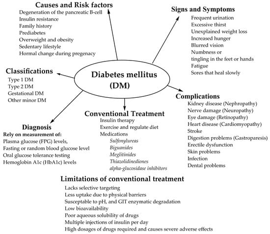
15 pages, 3746 KiB  
Review
Shining Light on Protein Kinase Biomarkers with Fluorescent Peptide Biosensors
by May C. Morris
Life 2022, 12(4), 516; https://doi.org/10.3390/life12040516 - 31 Mar 2022
Cited by 4 | Viewed by 2911
Abstract
Protein kinases (PKs) are established gameplayers in biological signalling pathways, and a large body of evidence points to their dysregulation in diseases, in particular cancer, where rewiring of PK networks occurs frequently. Fluorescent biosensors constitute attractive tools for probing biomolecules and monitoring dynamic [...] Read more.
Protein kinases (PKs) are established gameplayers in biological signalling pathways, and a large body of evidence points to their dysregulation in diseases, in particular cancer, where rewiring of PK networks occurs frequently. Fluorescent biosensors constitute attractive tools for probing biomolecules and monitoring dynamic processes in complex samples. A wide variety of genetically encoded and synthetic biosensors have been tailored to report on PK activities over the last decade, enabling interrogation of their function and insight into their behaviour in physiopathological settings. These optical tools can further be used to highlight enzymatic alterations associated with the disease, thereby providing precious functional information which cannot be obtained through conventional genetic, transcriptomic or proteomic approaches. This review focuses on fluorescent peptide biosensors, recent developments and strategies that make them attractive tools to profile PK activities for biomedical and diagnostic purposes, as well as insights into the challenges and opportunities brought by this unique toolbox of chemical probes. Full article
Show Figures

Figure 1

Figure 1
<p>Complexity of the protein kinase activity/function illustrated by the RAS/RAF/MEK/ERK/CDK4 pathway. (<b>a</b>) PK activity is the result of a number of factors: CDK4 expression level, its regulation by activating and inhibitory partners, such as cyclin D and structural inhibitor p16INK4, posttranslational modifications catalysed by upstream regulators, such as ERK, MEK, RAS and RAF. (<b>b</b>) In human cancers, CDK4 hyperactivity may occur through different mechanisms that affect CDK4, cyclin D or p16INK4.</p>
Full article ">Figure 2
<p>Strategies to detect and quantify PK expression and function: (<b>a</b>) genetic profiling; (<b>b</b>) transcriptomic profiling; (<b>c</b>) proteomics and interaction networks based on mass spectrometry analyses; (<b>d</b>) antibody-based approaches (ELISA and Western blotting); (<b>e</b>) methods based on ATP labelling; (<b>f</b>) fluorescent biosensors—kinase activity reporters.</p>
Full article ">Figure 3
<p>Toolbox and diversity of fluorescent biosensors. (<b>a</b>) Genetically encoded FRET kinase activity reporter; (<b>b</b>) synthetic peptide biosensors; (<b>c</b>) nanomaterial-based biosensors: single-wall and multiwall carbon nanotube biosensors (<b>left</b> and <b>middle</b>, respectively) and a graphene biosensor with antibodies and fluorescent enzymes (<b>right</b>).</p>
Full article ">Figure 4
<p>Features characterizing performance and robustness of fluorescent biosensors.</p>
Full article ">Figure 5
<p>Mechanisms of response of fluorescent peptide biosensors. (<b>a</b>) Phosphorylation of the peptide affects fluorescence emission of the fluorophore proximal to the phosphate group; (<b>b</b>) phosphorylation of the peptide substrate promotes binding of a phosphoamino acid-binding domain which alters the local environment; (<b>c</b>) a quencher proximal to the fluorophore reduces its basal fluorescence until phosphorylation disrupts quenching; (<b>d</b>) chelation-enhanced fluorescence of a fluorophore upon binding of a metal ion involved in the phosphorylation process; (<b>e</b>,<b>f</b>) aggregation-caused quenching of fluorophores is reversed upon phosphorylation which promotes disassembly of fluorescent biosensors, leading to fluorescence enhancement.</p>
Full article ">Figure 6
<p>CDKACT fluorescent peptide biosensor technology. (<b>a</b>) Schematic representation of CDKACT biosensors: bipartite biosensors comprising a CDK-specific substrate moiety (orange) onto which an environmentally sensitive dye is conjugated, a short linker and a phosphoamino acid-binding domain (or PAABD, green) that folds onto the phosphorylated substrate, thereby altering the local environment of the fluorophore and promoting changes in fluorescence emission. (<b>b</b>) An RFP fusion of CDKACT expressed in <span class="html-italic">E. coli</span>. (<b>c</b>) CDKACTs or RFP-CDKACTs can be introduced into cultured cells for live imaging experiments through complexation with cell-penetrating peptides (Pep1) to form nanoparticles that cross cell membranes and release CDKACT into cells. (<b>d</b>) A self-cell-penetrating variant of CDKACT was generated through fusion of the N-terminal moiety of Pep1 to the PAABD of CDKACT5. (<b>e</b>) CDKACT1-multiwall carbon nanotube conjugates yield an ultrasensitive nano-biosensor for imaging the CDK1 activity in mice.</p>
Full article ">Figure 7
<p>CDKACT fluorescent peptide biosensor technology. (<b>a</b>) CDKACT technology for quantifying the CDK activity in vitro by means of fluorescence spectroscopy and in living cells by means of fluorescence microscopy and live cell imaging following facilitated delivery into cultured cells. (<b>b</b>) Comparison of fluorescent and luminescent CDKACT4 biosensors. (<b>c</b>) Schematic representation of multiplex biosensing experiments performed by combining four different CDKACT biosensors to profile CDK1, CDK2, CDK4 and CDK6 in human cancer biopsies.</p>
Full article ">
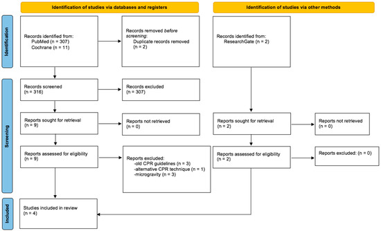
24 pages, 6644 KiB  
Review
Next-Generation Molecular Discovery: From Bottom-Up In Vivo and In Vitro Approaches to In Silico Top-Down Approaches for Therapeutics Neogenesis
by Sophie E. Kenny, Fiach Antaw, Warwick J. Locke, Christopher B. Howard, Darren Korbie and Matt Trau
Life 2022, 12(3), 363; https://doi.org/10.3390/life12030363 - 2 Mar 2022
Cited by 2 | Viewed by 6511
Abstract
Protein and drug engineering comprises a major part of the medical and research industries, and yet approaches to discovering and understanding therapeutic molecular interactions in biological systems rely on trial and error. The general approach to molecular discovery involves screening large libraries of [...] Read more.
Protein and drug engineering comprises a major part of the medical and research industries, and yet approaches to discovering and understanding therapeutic molecular interactions in biological systems rely on trial and error. The general approach to molecular discovery involves screening large libraries of compounds, proteins, or antibodies, or in vivo antibody generation, which could be considered “bottom-up” approaches to therapeutic discovery. In these bottom-up approaches, a minimal amount is known about the therapeutics at the start of the process, but through meticulous and exhaustive laboratory work, the molecule is characterised in detail. In contrast, the advent of “big data” and access to extensive online databases and machine learning technologies offers promising new avenues to understanding molecular interactions. Artificial intelligence (AI) now has the potential to predict protein structure at an unprecedented accuracy using only the genetic sequence. This predictive approach to characterising molecular structure—when accompanied by high-quality experimental data for model training—has the capacity to invert the process of molecular discovery and characterisation. The process has potential to be transformed into a top-down approach, where new molecules can be designed directly based on the structure of a target and the desired function, rather than performing screening of large libraries of molecular variants. This paper will provide a brief evaluation of bottom-up approaches to discovering and characterising biological molecules and will discuss recent advances towards developing top-down approaches and the prospects of this. Full article
Show Figures

Figure 1

Figure 1
<p>Bottom-up and top-down approaches to ex vivo molecular discovery. Bottom-up approaches generally involve screening broad libraries of candidate molecules against a target of interest and narrowing this down to a single best binder. The top-down approach is to design and generate new binders directly based on the structure of a target molecule.</p>
Full article ">Figure 2
<p>The current journey of therapeutics from discovery to the clinic (top to bottom), with prominent examples given of in vivo, in vitro and in silico approaches. A broad approximation of time taken for each stage of development is provided. (Created with <a href="http://lucidchart.com" target="_blank">lucidchart.com</a>).</p>
Full article ">Figure 3
<p>Traditional process of in vivo antibody isolation. First, animals are immunised with an antigen of interest. After an immune response has been mounted, B cells are isolated from the spleen of the animal and fused with an immortal myeloma cell line. Hybridomas are selected and screened for their activity against the antigen of interest. Target-binding clones are retained and used to obtain purified antibodies.</p>
Full article ">Figure 4
<p>Traditional library biopanning and enrichment against a target. Broadly, the genetically encoded library is incubated against a surface-bound antigen—the biological target of choice. Unbound phage are washed away whilst target-bound phage are reserved. Target-bound phage are then eluted from the immobilised antigen and regrown in <span class="html-italic">E. coli</span>. The now affinity-enriched phage cohort is then fed into the next cycle of biopanning.</p>
Full article ">Figure 5
<p>Some of the major advantages and disadvantages to each broad approach to therapeutics discovery, with the trend moving towards increased use of in silico technologies.</p>
Full article ">
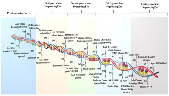
18 pages, 3384 KiB  
Hypothesis
Potassium at the Origins of Life: Did Biology Emerge from Biotite in Micaceous Clay?
by Helen Greenwood Hansma
Life 2022, 12(2), 301; https://doi.org/10.3390/life12020301 - 17 Feb 2022
Cited by 11 | Viewed by 4952 | Correction
Abstract
Intracellular potassium concentrations, [K+], are high in all types of living cells, but the origins of this K+ are unknown. The simplest hypothesis is that life emerged in an environment that was high in K+. One such environment [...] Read more.
Intracellular potassium concentrations, [K+], are high in all types of living cells, but the origins of this K+ are unknown. The simplest hypothesis is that life emerged in an environment that was high in K+. One such environment is the spaces between the sheets of the clay mineral mica. The best mica for life’s origins is the black mica, biotite, because it has a high content of Mg++ and because it has iron in various oxidation states. Life also has many of the characteristics of the environment between mica sheets, giving further support for the possibility that mica was the substrate on and within which life emerged. Here, a scenario for life’s origins is presented, in which the necessary processes and components for life arise in niches between mica sheets; vesicle membranes encapsulate these processes and components; the resulting vesicles fuse, forming protocells; and eventually, all of the necessary components and processes are encapsulated within individual cells, some of which survive to seed the early Earth with life. This paper presents three new foci for the hypothesis of life’s origins between mica sheets: (1) that potassium is essential for life’s origins on Earth; (2) that biotite mica has advantages over muscovite mica; and (3) that micaceous clay is a better environment than isolated mica for life’s origins. Full article
Show Figures

Figure 1

Figure 1
<p>[K<sup>+</sup>] between mica sheets. Structure of the black mica, biotite. (<b>A</b>) Side view of three biotite sheets, labeled ‘1’, ‘2’ and ‘3’. (<b>B</b>) Top view of 1 nm<sup>2</sup> biotite, with K<sup>+</sup> highlighted in the right-hand image, showing that there are six K<sup>+</sup> per nm<sup>2</sup> between mica sheets. (<b>C</b>) Side view of six mica sheets that are not separated and separated at a distance of 1 nm, where [K<sup>+</sup>] = 10 M between the sheets. (<b>D</b>) Scale model of biotite sheets at a separation of 100 nm, where [K<sup>+</sup>] = 100 mM. (CrystalMaker X software, version 10.6.4, CrystalMaker Software Ltd., Oxfordshire, UK).</p>
Full article ">Figure 2
<p>Nanometer-scale diagram of how the early stages of life might have originated between biotite mica sheets. Niches within the biotite sheets provide partially enclosed spaces for the molecular evolution of the different processes that are essential for life. Vesicles form, encapsulating molecules and molecular complexes from the niches.</p>
Full article ">Figure 3
<p>Micron-scale diagram of how life might have originated between biotite mica sheets. Protocells in the aqueous environment encapsulate prebiotic molecular aggregates in the niches between mica sheets. Mechanical energy from moving mica sheets can bleb off protocells, as seen in the lower left corner of the figure. Eventually, a living cell capable of self-reproduction will eventually be produced.</p>
Full article ">Figure 4
<p>Mica and mechanical energy. (<b>A</b>) Diagram of mechanical forces between biotite mica sheets stretching and compressing polymers due to water flow at the edges of the biotite sheets. (<b>B</b>) Diagram of mechanical forces between biotite mica sheets due to heat pumps in a biotite bubble. This mechanical energy can be used to synthesize prebiotic molecules, stretch and compress polymers (as shown in the diagram), or bleb off protocells [<a href="#B23-life-12-00301" class="html-bibr">23</a>]. Seven mica sheets, as shown in (<b>A</b>), provide enough force to form a covalent bond in air when moved to a distance of 0.1 nm. (<b>C</b>) Biotite bubble (arrows) imaged by HRTEM (high-resolution transmission electron microscopy) [<a href="#B65-life-12-00301" class="html-bibr">65</a>] with expanded view of HRTEM image and CrystalMaker model of biotite on left. “{“ or “}o” = two biotite layers. The thickness of a single biotite sheet is 1 nm (10 Angstroms). (<b>D</b>) Photograph of muscovite mica, showing a bubble (upper arrow) and separation at the edges of the mica sheet (lower arrow). Bubbles are common, even in “high grade” micas such as this one.</p>
Full article ">Figure 5
<p>Micaceous clay and the origin of life between mica sheets. (<b>A</b>) Mica origins diagram of <a href="#life-12-00301-f003" class="html-fig">Figure 3</a>, a late stage in the origin of life, extended to a length of 80 microns. (<b>B</b>) Mica Red Micaceous Clay from a New Mexico Clay Store containing pale reflecting pieces of mica in the middle of a wet-dry cycle. (<b>C</b>) Mica and a few clay particles washed from the micaceous clay. Yellow asterisk and arrow point to a mica fragment with a diameter of ~80 microns. Yellow asterisks in (<b>A</b>,<b>C</b>) indicate mica fragments of ~80-microns.</p>
Full article ">Figure 6
<p>Swelling clay (montmorillonite) and non-swelling clays (muscovite and biotite). Sodium ions, Na<sup>+</sup>, (mustard color) bridge sheets of montmorillonite (mustard-colored arrows); potassium ions, K<sup>+</sup>, (purple) bridge sheets of muscovite and biotite (purple arrows). Ionic radii are 0.095 nm for Na<sup>+</sup> and 0.133 nm for K<sup>+</sup> [<a href="#B90-life-12-00301" class="html-bibr">90</a>]. The smaller Na<sup>+</sup> are hydrated between montmorillonite sheets, which causes montmorillonite to swell and shrink with wetting and drying. The larger K<sup>+</sup> between biotite sheets are not hydrated; biotite does not swell and shrink with wetting and drying. Molecular models show two sheets of muscovite mica (left), montmorillonite clay (center), biotite mica (right), and water molecules. Surfaces of the sheets of all three clay crystals are tetrahedral silicon–oxygen (Si-O) layers; see <a href="#life-12-00301-f001" class="html-fig">Figure 1</a>B for top view. Clay models and water molecules show relative sizes of atoms and structures. Atom identification list on the right does not show relative sizes of atoms. Atom list = K, Na, Si, Al, Mg, Fe, H, O, OH. (CrystalMaker X software, version 10.6.4, CrystalMaker Software Ltd., Oxfordshire, UK).</p>
Full article ">Figure 7
<p>Atomic Force Microscopy of circular double-stranded DNA (dsDNA) on mica with cracks. (Cracks are dark streaks at the upper left). Three of the four dsDNA circles formed a 60<sup>o</sup> angle with the mica crack, consistent with the alignment on the mica’s hexagonal crystal lattice.</p>
Full article ">
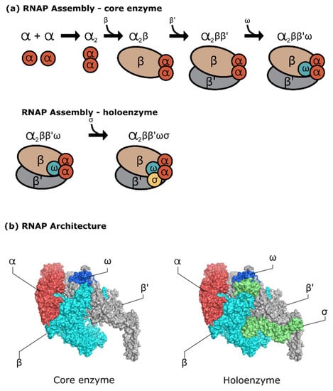
16 pages, 795 KiB  
Review
Of Cockroaches and Symbionts: Recent Advances in the Characterization of the Relationship between Blattella germanica and Its Dual Symbiotic System
by Amparo Latorre, Rebeca Domínguez-Santos, Carlos García-Ferris and Rosario Gil
Life 2022, 12(2), 290; https://doi.org/10.3390/life12020290 - 15 Feb 2022
Cited by 6 | Viewed by 4901
Abstract
Mutualistic stable symbioses are widespread in all groups of eukaryotes, especially in insects, where symbionts have played an essential role in their evolution. Many insects live in obligate relationship with different ecto- and endosymbiotic bacteria, which are needed to maintain their hosts’ fitness [...] Read more.
Mutualistic stable symbioses are widespread in all groups of eukaryotes, especially in insects, where symbionts have played an essential role in their evolution. Many insects live in obligate relationship with different ecto- and endosymbiotic bacteria, which are needed to maintain their hosts’ fitness in their natural environment, to the point of even relying on them for survival. The case of cockroaches (Blattodea) is paradigmatic, as both symbiotic systems coexist in the same organism in two separated compartments: an intracellular endosymbiont (Blattabacterium) inside bacteriocytes located in the fat body, and a rich and complex microbiota in the hindgut. The German cockroach Blattella germanica is a good model for the study of symbiotic interactions, as it can be maintained in the laboratory in controlled populations, allowing the perturbations of the two symbiotic systems in order to study the communication and integration of the tripartite organization of the host–endosymbiont–microbiota, and to evaluate the role of symbiotic antimicrobial peptides (AMPs) in host control over their symbionts. The importance of cockroaches as reservoirs and transmission vectors of antibiotic resistance sequences, and their putative interest to search for AMPs to deal with the problem, is also discussed. Full article
Show Figures

Figure 1

Figure 1
<p>Life cycle of <span class="html-italic">B. germanica</span> and experimental design for antibiotic treatment and recovery. (<b>a</b>) The life cycle of <span class="html-italic">B. germanica</span> consists of three developmental stages: egg, nymph (N) and adult (A), and is completed in approximately 95 days at 26 °C. The egg begins its development inside the ootheca (egg case), which contains around 30–40 eggs and remains attached to the female until the nymphs are born. After approximately 35 days, the nymphs emerge from the ootheca (ootheca hatching, d0N). Nymphs moult and go through 5 or 6 nymphal stages (N1 through N6), shedding their exoskeleton and growing, and reach full maturity in approximately 50 days (d50N). After the final moulting (adult ecdysis), the sexually active adult emerges (stage with wings, d0A). Adults breed immediately, and the ootheca with fertilized eggs emerge in the female in just one week. (<b>b</b>) A typical experiment starts with a synchronized adult population, composed of individuals collected between 0 and 48 h after adult ecdysis in generation 1 (G1). Then, the population is divided into two subpopulations: the control one is never treated with antibiotics (−AB), until reaching the second generation (G2); the experimental one is treated with antibiotics (+AB) during the adult stage. The antibiotics are removed when the ootheca is fully formed (a few days before hatching), and newly emerged nymphs are divided into three subpopulations in G2: the first one is immediately treated with antibiotics (+AB) during the complete life cycle (Treatment), the second one is kept without any further antibiotic treatment (−AB; Recovery) and the third one is kept without any further antibiotic treatment, but is supplemented with faeces obtained from a control population that has been never treated with the antibiotic (−AB+F; Recovery with faeces).</p>
Full article ">
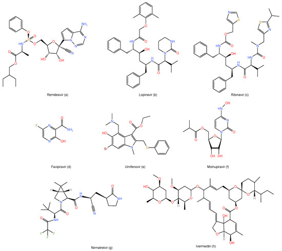
16 pages, 82661 KiB  
Review
Were the First Trace Fossils Really Burrows or Could They Have Been Made by Sediment-Displacive Chemosymbiotic Organisms?
by Duncan McIlroy
Life 2022, 12(2), 136; https://doi.org/10.3390/life12020136 - 18 Jan 2022
Cited by 3 | Viewed by 4157
Abstract
This review asks some hard questions about what the enigmatic graphoglyptid trace fossils are, documents some of their early fossil record from the Ediacaran–Cambrian transition and explores the idea that they may not have been fossils at all. Most researchers have considered the [...] Read more.
This review asks some hard questions about what the enigmatic graphoglyptid trace fossils are, documents some of their early fossil record from the Ediacaran–Cambrian transition and explores the idea that they may not have been fossils at all. Most researchers have considered the Graphoglyptida to have had a microbial-farming mode of life similar to that proposed for the fractal Ediacaran Rangeomorpha. This begs the question “What are the Graphoglyptida if not the Rangeomorpha persevering” and if so then “What if…?”. This provocative idea has at its roots some fundamental questions about how to distinguish burrows sensu-stricto from the external molds of endobenthic sediment displacive organisms. Full article
Show Figures

Figure 1

Figure 1
<p>(<b>A</b>) Long, narrow Ediacaran frond from Mistaken Point Ecological reserve, NL (scale bar in mm); (<b>B</b>) large reclining rangeomorph Ediacaran frond. aff. <span class="html-italic">Bradgatia</span> sp. from the MUN surface, Catalina Dome, NL; (<b>C</b>) diagrammatic reconstruction of a generic reclining rangeomorph detailing the ways that it might have interacted with the substrate. The lower surface is irrigated with seawater by ciliary action and diffusion. The supply of oxygen to the lower surface is considered to have increased microbial productivity. The top row of images shows possible feeding modes with green circles showing the distribution of chemolithoautotrophic symbionts and arrows show diffusion of solutes. Furthest left is phagotrophy, next is ectosymbiosis, then endosymbiosis and furthest right is endosymbiosis with a trophosome (requiring diffusion of sulfide/methane into a thin organism and POM = particulate organic matter). All of these methods of gaining nutrition would work for endobenthic graphoglyptids; (<b>D</b>) reconstruction of the Ediacaran seafloor of Mistaken Point Formation (courtesy of Paleocreations).</p>
Full article ">Figure 2
<p>(<b>A</b>) Microbial matground surface with wrinkled texture and abundant sediment mining trace fossils from the Ordovician of Bell Island, NL; (<b>B</b>) typical ichnofabric from the lower Fortunian of Fortune Head showing abundant curved, spiraling and branching pyritized burrows; (<b>C</b>,<b>D</b>) bedding plane view of <span class="html-italic">Lamonte trevallis</span> burrows (Lt) and pyritized graphoglyptid burrows (PGr) with T junction arrowed. Scale bars 1 cm.</p>
Full article ">Figure 3
<p>Tubular open “burrows” from the Fortunian of Fortune Head NL showing spiraling morphologies of: (<b>A</b>) <span class="html-italic">Gyrolithes gyratus</span>; and (<b>B</b>) <span class="html-italic">G. scintillus</span> with pyrite rich silty sandstone fill. (<b>C</b>) Shows the bedding plane view of a uniserially branching <span class="html-italic">Treptichnus pedum</span> in which the pyritic fill has weathered away showing the mold of the burrow, the space that would have been occupied in life. Whether these structures were burrows sensu-stricto or casts of the exterior of spiraling or branching organisms remains to be determined. (<b>D</b>) Natural sandstone cast of <span class="html-italic">T. pedum</span> from the Arumbera Formation in conventional positive hyporelief Scale bars 1 cm.</p>
Full article ">Figure 4
<p>Segments of <span class="html-italic">Trichophycus</span> ispp. from the lower Cambrian Arumbera Sandstone of central Australia showing the stacked spreite (arrowed bottom left) and scratch marks (arrowed top right) that distinguish the genus from <span class="html-italic">Treptichnus</span>. Scale bar 1 cm.</p>
Full article ">Figure 5
<p>Graphoglyptid morphologies: (<b>A</b>,<b>B</b>) are line graphoglyptids ((<b>A</b>) <span class="html-italic">Helminthoida</span> though note the branching from the Cambrian Arumbera Sandstone Australia; (<b>B</b>) is <span class="html-italic">Helicolithus</span> from the latest Ediacaran of Tanafjord, Norway); (<b>C</b>,<b>D</b>) are branching graphoglytids. ((<b>C</b>) is <span class="html-italic">Belorhaphe</span> from the late Ediacaran of Tanafjord, Norway, (<b>D</b>) is cf. <span class="html-italic">Paleomeandron</span> from the Cambrian Arumbera Sandstone); (<b>E</b>,<b>F</b>) are net graphoglyptids ((<b>E</b>) is <span class="html-italic">Squamodictyon</span> and (<b>F</b>) is <span class="html-italic">Paleodictyon</span> from the Arumbera Sandstone, Australia). Scale bars 1 cm.</p>
Full article ">Figure 6
<p>(<b>A</b>–<b>D</b>) <span class="html-italic">Treptichnus pedum</span> showing a range of branching types. All preserved as open burrow fills from the Fortunian of Tanafjord, Norway. D shows both uniserial and biserial monopodial branching. (<b>E</b>) is a small portion of aff. <span class="html-italic">Bradgatia</span> showing <span class="html-italic">Treptichnus</span> such as branching. (<b>F</b>) <span class="html-italic">Treptichnus lublinensis</span> showing meandering habit and very rangeomorph-like branching. Scale bars 1 cm except (<b>D</b>), which is 5 cm.</p>
Full article ">Figure 7
<p>Net-type graphoglyptids from the Ediacaran (<b>A</b>) <span class="html-italic">Multina</span> and Fortunian (<b>B</b>) <span class="html-italic">Paleodictyon</span> of Tanafjord. Some of the supposedly most complicated marine trace fossils amidst the earliest record of endobenthic activity. Pen for scale in (<b>A</b>) is 1 cm diameter, scale bar in (<b>B</b>) 1 cm.</p>
Full article ">
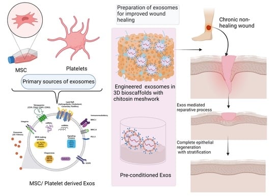
2021

Jump to: 2024, 2023, 2022

34 pages, 1112 KiB  
Review
Endothelial Dysfunction in Childhood Cancer Survivors: A Narrative Review
by Marco Crocco, Giuseppe d’Annunzio, Alberto La Valle, Gianluca Piccolo, Decimo Silvio Chiarenza, Carolina Bigatti, Marta Molteni, Claudia Milanaccio, Maria Luisa Garrè, Natascia Di Iorgi and Mohamad Maghnie
Life 2022, 12(1), 45; https://doi.org/10.3390/life12010045 - 29 Dec 2021
Cited by 4 | Viewed by 3181
Abstract
Assessment of endothelial dysfunction in cancer survivors may have a role in the early identification of non-communicable diseases and cardiovascular late effects. Oncological therapies may impair endothelial function. Therefore, in patients such as childhood cancer survivors who could benefit from early cardioprotective pharmacological [...] Read more.
Assessment of endothelial dysfunction in cancer survivors may have a role in the early identification of non-communicable diseases and cardiovascular late effects. Oncological therapies may impair endothelial function. Therefore, in patients such as childhood cancer survivors who could benefit from early cardioprotective pharmacological interventions, it is essential to monitor endothelial function, even if the optimal methodology for investigating the multifaceted aspects of endothelial dysfunction is still under debate. Biochemical markers, as well as invasive and non-invasive tools with and without pharmacological stimuli have been studied. Human clinical studies that have examined lifestyle or cancer treatment protocols have yielded evidence showing the involvement of lipid and lipoprotein levels, glycemic control, blood pressure, adiposity, inflammation, and oxidative stress markers on the state of endothelial health and its role as an early indicator of cardiometabolic risk. However, with regards to pharmacological interventions, cautious interpretation of the result attained whilst monitoring the endothelial function is warranted due to methodological limitations and substantial heterogeneity of the results reported in the published studies. In this narrative review, an overview of evidence from human clinical trials examining the effects of cancer therapies on endothelial disease is provided together with a discussion of endothelial function assessment using the different non-invasive techniques available for researchers and clinicians, in recent years. Full article
Show Figures

Figure 1

Figure 1
<p>Flow chart for eligible studies. * letters to editors, book chapters, study protocols, case reports.</p>
Full article ">Figure 2
<p>Etiopathogenesis of endothelial dysfunction in cancer survivors.</p>
Full article ">
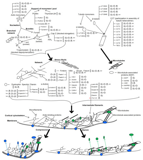
19 pages, 1427 KiB  
Review
From Life in the Sea to the Clinic: The Marine Drugs Approved and under Clinical Trial
by Emiliano Cappello and Paola Nieri
Life 2021, 11(12), 1390; https://doi.org/10.3390/life11121390 - 11 Dec 2021
Cited by 33 | Viewed by 6593
Abstract
In the last decades Blue Growth policy in european and non-european countries produced a great impulse in applied marine sciences, comprehending the research of new bioactive molecules in marine organisms. These organisms are a great source of natural compounds with unique features resulting [...] Read more.
In the last decades Blue Growth policy in european and non-european countries produced a great impulse in applied marine sciences, comprehending the research of new bioactive molecules in marine organisms. These organisms are a great source of natural compounds with unique features resulting from the huge variability of marine habitats and species living in them. Most of the marine compounds in use and in clinical trials are drugs for cancer therapy and many of them are conjugated to antibody to form antibody-drug conjugates (ADCs). Severe pain, viral infections, hypertriglyceridemia, obesity, Alzheimer’s and other CNS diseases are further target conditions for these pharmaceuticals. This review summarizes the state-of-the-art marine drugs focusing on the most successful results in the fast expanding field of marine pharmacology. Full article
Show Figures

Graphical abstract

Graphical abstract
Full article ">Figure 1
<p>Chemical structures of marine drugs approved or under clinical trials, not including those present in antibody drug conjugates (ADCs) which are reported in <a href="#life-11-01390-f002" class="html-fig">Figure 2</a>.</p>
Full article ">Figure 2
<p>(<b>A</b>) General structure of Antibody-Drug Conjugates (ADCs) approved and in clinical trials and (<b>B</b>) chemical structures of toxins.</p>
Full article ">Figure 3
<p>(<b>A</b>) Number of marine drugs under clinical trials and already marketed (in use), distributed for therapeutic class; (<b>B</b>) % distribution of all marine drugs (under clinical trials and already marketed) for their use (cancer vs. non cancer); (<b>C</b>) % distribution of marine anticancer agents (under clinical trials and already marketed) in different typology.</p>
Full article ">
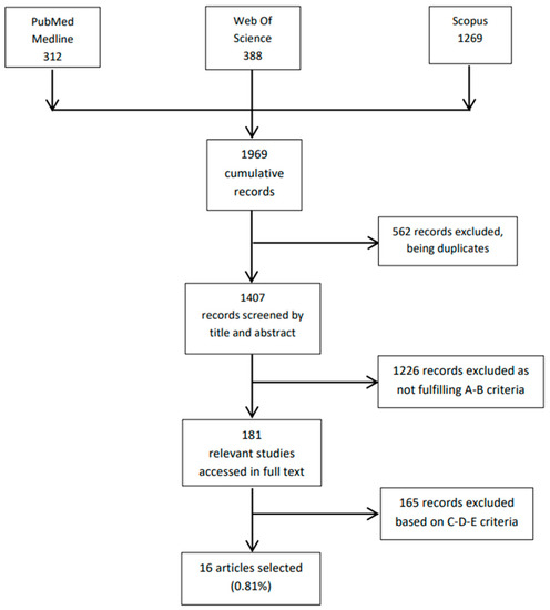
17 pages, 1094 KiB  
Review
Potential Reasons for Unresponsiveness to Anti-PD1 Immunotherapy in Young Patients with Advanced Melanoma
by Devayani Machiraju, Sarah Schäfer and Jessica C. Hassel
Life 2021, 11(12), 1318; https://doi.org/10.3390/life11121318 - 30 Nov 2021
Cited by 12 | Viewed by 4784
Abstract
The impact of age on the clinical benefit of anti-PD1 immunotherapy in advanced melanoma patients has been evolving recently. Due to a reduced immune function in elderly patients, young patients with a robust immune system are theoretically expected to benefit more from the [...] Read more.
The impact of age on the clinical benefit of anti-PD1 immunotherapy in advanced melanoma patients has been evolving recently. Due to a reduced immune function in elderly patients, young patients with a robust immune system are theoretically expected to benefit more from the treatment approach. However, in contrast to this hypothesis, recent studies in patients with metastatic melanoma have demonstrated that immunotherapy, especially with anti-PD1 treatment, is less effective in patients below 65 years, on average, with significantly lower responses and reduced overall survival compared to patients above 65 years of age. Besides, data on young patients are even more sparse. Hence, in this review, we will focus on age-dependent differences in the previously described resistance mechanisms to the treatment and discuss the development of potential combination treatment strategies for enhancing the anti-tumor efficacy of anti-PD1 or PDL1 treatment in young melanoma patients. Full article
Show Figures

Figure 1

Figure 1
<p>Age-related differences in clinical and molecular factors associated with anti-PD1 treatment outcome in metastatic melanoma patients.</p>
Full article ">Figure 2
<p>Potential combination treatments to enhance anti-PD1 treatment efficacy in younger patients with metastatic melanoma.</p>
Full article ">
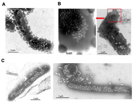
19 pages, 31908 KiB  
Review
Automated Exploration of Prebiotic Chemical Reaction Space: Progress and Perspectives
by Siddhant Sharma, Aayush Arya, Romulo Cruz and Henderson James Cleaves II
Life 2021, 11(11), 1140; https://doi.org/10.3390/life11111140 - 26 Oct 2021
Cited by 6 | Viewed by 7819
Abstract
Prebiotic chemistry often involves the study of complex systems of chemical reactions that form large networks with a large number of diverse species. Such complex systems may have given rise to emergent phenomena that ultimately led to the origin of life on Earth. [...] Read more.
Prebiotic chemistry often involves the study of complex systems of chemical reactions that form large networks with a large number of diverse species. Such complex systems may have given rise to emergent phenomena that ultimately led to the origin of life on Earth. The environmental conditions and processes involved in this emergence may not be fully recapitulable, making it difficult for experimentalists to study prebiotic systems in laboratory simulations. Computational chemistry offers efficient ways to study such chemical systems and identify the ones most likely to display complex properties associated with life. Here, we review tools and techniques for modelling prebiotic chemical reaction networks and outline possible ways to identify self-replicating features that are central to many origin-of-life models. Full article
Show Figures

Figure 1

Figure 1
<p>Some of the current questions and methods for the exploration of prebiotic CRNRs.</p>
Full article ">Figure 2
<p>CRNRs may accurately predict the many features of CRNs. (<b>A</b>) The 150–210 amu region of the mass spectrum of the products of a laboratory formose reaction measured using high-resolution Fourier transform ion cyclotron resonance mass spectrometry (FT-ICR-MS) in negative ESI mode. (<b>B</b>) The predicted mass distribution after six generations of the products of the same reaction was generated using CRNR methods. In this formose reaction, 2 M paraformaldehyde, 1 M glycolaldehyde, and 0.05 M Ca(OH)<math display="inline"><semantics> <msub> <mrow/> <mn>2</mn> </msub> </semantics></math> were heated in aqueous solution in sealed glass ampoules under nitrogen at 85° for eight days. The code for generating this figure is described in <a href="#app1-life-11-01140" class="html-app">Supplementary Materials</a>.</p>
Full article ">Figure 3
<p>Schematic for the forward synthesis of a network using graph theory-based tools. Typically, a set of “reaction rules” are loaded, which specify how certain substructures are to be altered during a reaction. All the rules are then applied combinatorially to a set of initial reactants, which gives a set of products as the graph transformations happen that give product molecules, connected by “edges” representing reactions. Constraints that certain molecules (or “graphs”) with certain substructures should not form can be used to filter out unstable species. Iterative application of these rules to the product suite at each step gives a complete CRNR.</p>
Full article ">Figure 4
<p>A primitive CRNR for glucose degradation reaction, generated using the graph grammar-based program MØD. The in silico synthesis of this CRNR was done as per the general methods outlined in <a href="#life-11-01140-f003" class="html-fig">Figure 3</a>. The reaction rules used were selected based on prior knowledge of mechanisms known to dominate in this chemical system under pH and temperature conditions of interest. A single cycle of reaction rule application (i.e., one “generation”) is shown here. In the visual representation used here, molecules are shown in ovals, while reaction nodes are shown as squares.</p>
Full article ">Figure 5
<p>General features of autocatalysis and a specific example of an autocatalytic cycle detectable within CRNRs. (<b>A</b>) The basic idea of autocatalysis is that a sequence (or network) of reactions begins with a specific molecule A and produces more than one copy of A, assuring that the cycle produces more A than it consumes. (<b>B</b>) A concrete example of such a cycle within a larger CRNR. Here, two paths, <math display="inline"><semantics> <mrow> <mi>A</mi> <mo>→</mo> <mi>B</mi> <mo>→</mo> <mi>E</mi> <mo>→</mo> <mi>F</mi> <mo>→</mo> <mi>A</mi> </mrow> </semantics></math> and <math display="inline"><semantics> <mrow> <mi>A</mi> <mo>→</mo> <mi>B</mi> <mo>→</mo> <mi>C</mi> <mo>→</mo> <mi>D</mi> <mo>→</mo> <mi>A</mi> </mrow> </semantics></math>, contribute to produce stoichiometrically larger quantities of A. The CRNR illustrated here was produced using five rounds of reaction generation in the glucose degradation chemistry discussed above. The layout of the graph was executed using Gephi. The size of the nodes corresponds to each node in-degree. Each color represents a new generation.</p>
Full article ">Figure 6
<p>Gephi representations of CRNRs for (<b>A</b>) glucose degradation, (<b>B</b>) formose reaction, (<b>C</b>) pyruvic acid degradation, and (<b>D</b>) the reaction of HCN + NH<sub>3</sub>. All CRNRs were generated using the graph-grammar techniques previously discussed, and the figures shown here were produced using the Gephi software. Such visualization tools are helpful for broad visual classification of CRNRs. The size of the nodes in these graphs is proportional to the node’s in-degree as a function of how many edges reach it. Node color indicates the generation in which each compound was first generated. The code for generating this figure is described in <a href="#app1-life-11-01140" class="html-app">Supplementary Materials</a>.</p>
Full article ">
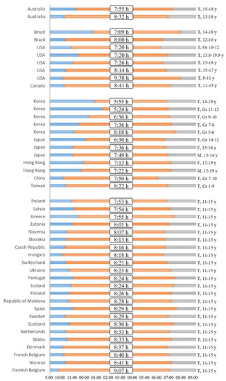
13 pages, 576 KiB  
Review
Uncovering the Roles of MicroRNAs in Major Depressive Disorder: From Candidate Diagnostic Biomarkers to Treatment Response Indicators
by Claudia Homorogan, Diana Nitusca, Edward Seclaman, Virgil Enatescu and Catalin Marian
Life 2021, 11(10), 1073; https://doi.org/10.3390/life11101073 - 11 Oct 2021
Cited by 13 | Viewed by 2688
Abstract
Major depressive disorder (MDD) is a recurrent debilitating illness that represents a major health burden due to its increasing worldwide prevalence, unclear pathological mechanism, nonresponsive treatment, and lack of reliable and specific diagnostic biomarkers. Recently, microRNA species (miRs) have gained particular interest because [...] Read more.
Major depressive disorder (MDD) is a recurrent debilitating illness that represents a major health burden due to its increasing worldwide prevalence, unclear pathological mechanism, nonresponsive treatment, and lack of reliable and specific diagnostic biomarkers. Recently, microRNA species (miRs) have gained particular interest because they have the ability to post-transcriptionally regulate gene expression by modulating mRNA stability and translation in a cohesive fashion. By regulating entire genetic circuitries, miRs have been shown to have dysregulated expression levels in blood samples from MDD patients, when compared to healthy subjects. In addition, antidepressant treatment (AD) also appears to alter the expression pattern of several miRs. Therefore, we critically and systematically reviewed herein the studies assessing the potential biomarker role of several candidate miRs for MDD, as well as treatment response monitoring indicators, in order to enrich the current knowledge and facilitate possible diagnostic biomarker development for MDD, which could aid in reducing both patients’ burden and open novel avenues toward a better understanding of MDD neurobiology. Full article
Show Figures

Figure 1

Figure 1
<p>Flow diagram of the study selection process.</p>
Full article ">
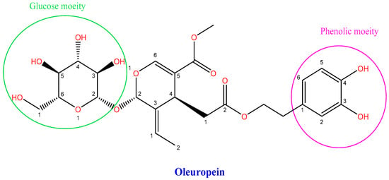
13 pages, 790 KiB  
Review
Understanding Vernal Keratoconjunctivitis: Beyond Allergic Mechanisms
by Marta Sacchetti, Rocco Plateroti, Alice Bruscolini, Rosalia Giustolisi and Marco Marenco
Life 2021, 11(10), 1012; https://doi.org/10.3390/life11101012 - 26 Sep 2021
Cited by 17 | Viewed by 5365
Abstract
Vernal keratoconjunctivitis (VKC) is a chronic, recurrent, inflammatory disease of the cornea and conjunctiva mostly affecting boys in prepubertal age. VKC recurrence is characterized by intense symptoms of itching, redness, and photophobia associated with corneal damage, impairment of visual function, and quality of [...] Read more.
Vernal keratoconjunctivitis (VKC) is a chronic, recurrent, inflammatory disease of the cornea and conjunctiva mostly affecting boys in prepubertal age. VKC recurrence is characterized by intense symptoms of itching, redness, and photophobia associated with corneal damage, impairment of visual function, and quality of life. The pathogenesis of VKC has not yet been completely understood, and it is still controversial. In fact, VKC is considered an ocular allergic disease due to the involvement of immunoglobulin E, eosinophils, and mast cells, and of a lymphocyte T-helper type 2 reaction. However, approximately half of VKC patients have negative allergological history and testing, suggesting that other pathogenic mechanisms participate in VKC development and severity. Specifically, evidence suggests that genetic, endocrine, neuronal factors and an imbalance of innate immunity are involved in the pathogenesis of VKC. The purpose of this review is to summarize evidence on the pathogenic role of innate immunity, neuroimmune reaction, and hormonal changes in VKC. Increasing understanding of the pathogenic mechanisms behind VKC may lead to the identification of novel biomarkers for diagnosis and/or potential therapeutic targets in order to improve the management of this challenging condition. Full article
Show Figures

Figure 1

Figure 1
<p>(<b>a</b>) Tarsal VKC with upper tarsal giant papillary reaction, (<b>b</b>) limbal VKC with limbal papillary reaction and the so-called Horner-Trantas dots.</p>
Full article ">Figure 2
<p>Corneal epithelial defect in a patient with VKC.</p>
Full article ">Figure 3
<p>Allergic reaction represents the main pathogenic factor of VKC; however, several sources of evidence show that innate immunity and neuroinflammatory response, as well as genetic, hormonal, and environmental factors, also participate in the development and severity of VKC.</p>
Full article ">
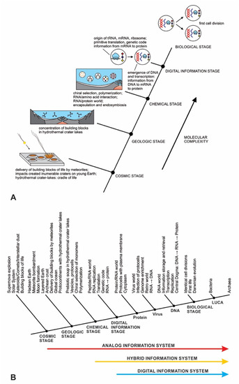
20 pages, 2931 KiB  
Review
The Epidemiology and Global Burden of Atopic Dermatitis: A Narrative Review
by Hazrina Ab Hadi, Aine Inani Tarmizi, Kamarul Ariffin Khalid, Márió Gajdács, Adeel Aslam and Shazia Jamshed
Life 2021, 11(9), 936; https://doi.org/10.3390/life11090936 - 9 Sep 2021
Cited by 97 | Viewed by 17573
Abstract
The global epidemiology of atopic dermatitis (AD) in the current decade (2009–2019) has not been extensively reported. Epidemiological studies play an important role in presenting the risk factors of AD, as detailed prevalence and incidence data could demonstrate the burden of disease in [...] Read more.
The global epidemiology of atopic dermatitis (AD) in the current decade (2009–2019) has not been extensively reported. Epidemiological studies play an important role in presenting the risk factors of AD, as detailed prevalence and incidence data could demonstrate the burden of disease in the population of adults, adolescents, and children in different geographical regions. Thus, the primary objective of this review was to assess and summarize the epidemiological studies of the prevalence and incidence of AD in different age groups, focusing on data from studies published for 2009 to 2019. After a thorough literature search, six countries were identified from African, Asian, and European regions respectively, who published studies on AD. In contrast, only two studies were identified from Australia and New Zealand, three countries from North America and two from South America published AD studies, respectively. The highest prevalence of AD from included studies was noted among Swedish children with 34%, while the lowest prevalence was in Tunisian children with 0.65%; studies reporting incidence data were far less numerous. A common trend in the prevalence of AD was that children would have a higher prevalence as compared to adolescents and adults. The severity and morbidity of the disease showed variance with age, sex, socioeconomic characteristics, geographical location, and ethnicity. Environmental factors played an important role as causative agents in AD. The risk factors that were proven to cause and induce AD were skin barrier impairments due to FLG mutation, changes in the environment, and diet. FLG mutation may impair the skin barrier function by disruption of pH and hydration maintenance of the skin. Lastly, there were only a few studies on the incidence of AD in the 21st century. Therefore, epidemiological studies on childhood and adulthood AD in different continents are still needed, especially on the incidence of AD during adulthood. Full article
Show Figures

Figure 1

Figure 1
<p>Chemical structures of the main components of the stratum corneum (SC) ceramides and their shorthand nomenclature.</p>
Full article ">Figure 2
<p>Graphic summary of effects of skin barrier on the pathogenesis of atopic dermatitis (AD). Genetic and immunologic as well as mechanical factors such as scratching induce skin barrier damage, allowing contact of skin resident antigen-presenting cells to allergens, bacterial, and viral antigens as well as other environmental factors. Activated antigen-presenting cells migrate to lymph nodes and prime naive T cells into Th2 cells. Elevated Th2 cytokines, together with TNF-α and IFN-γ further damage skin barrier functions by inducing apoptosis of keratinocytes as well as impair the function of tight junctions and promote Th2 responses by enhancing TSLP expression of epithelial cells. Moreover, colonizing pathogens, such as <span class="html-italic">Staphylococcus aureus</span>, impair barrier function through the release of virulence factors to induce keratinocyte death and to boost Th2-type inflammation. Together, genetic and immunological factors contribute to the skin barrier dysfunction and play a major role in the pathogenesis of AD.</p>
Full article ">Figure 3
<p>Prevalence of AD in Africa based on the reports published between 2009 and 2019.</p>
Full article ">Figure 4
<p>Prevalence of AD in America based on the reports published between 2009 and 2019.</p>
Full article ">Figure 5
<p>Prevalence of AD in Asia based on the reports published between 2009 and 2019.</p>
Full article ">Figure 6
<p>Prevalence of AD in Europe based on the reports published between 2009 and 2019.</p>
Full article ">Figure 7
<p>Prevalence of AD in Oceania based on the reports published between 2009 and 2019.</p>
Full article ">
12 pages, 1188 KiB  
Review
Horizontal Gene Transfers in Plants
by Emilie Aubin, Moaine El Baidouri and Olivier Panaud
Life 2021, 11(8), 857; https://doi.org/10.3390/life11080857 - 21 Aug 2021
Cited by 19 | Viewed by 6802
Abstract
In plants, as in all eukaryotes, the vertical transmission of genetic information through reproduction ensures the maintenance of the integrity of species. However, many reports over the past few years have clearly shown that horizontal gene transfers, referred to as HGTs (the interspecific [...] Read more.
In plants, as in all eukaryotes, the vertical transmission of genetic information through reproduction ensures the maintenance of the integrity of species. However, many reports over the past few years have clearly shown that horizontal gene transfers, referred to as HGTs (the interspecific transmission of genetic information across reproductive barriers) are very common in nature and concern all living organisms including plants. The advent of next-generation sequencing technologies (NGS) has opened new perspectives for the study of HGTs through comparative genomic approaches. In this review, we provide an up-to-date view of our current knowledge of HGTs in plants. Full article
Show Figures

Figure 1

Figure 1
<p>The three criteria used for HGT detection through comparative genomics: high similarity (HS), phylogenetic incongruence (PI) and patchy distribution in phylogenies (PD) [<a href="#B9-life-11-00857" class="html-bibr">9</a>]. The HS criterion requires access to genomic data of the two species involved in the transfer. It consists of establishing that gene homologs between two species exhibit a sequence identity that is significantly higher than the average of the other homologs in the genome, therefore not originating from vertical transmission. In order to circumvent the possible effect of strong selection (that could lead to high sequence identity), synonymous substitutions are used to measure sequence divergence. The PI criterion is based on the incongruence between the topologies of the phylogenetic tree of the species and that of the horizontally transferred genes. This requires sequence information for a large phylogenetic panel, as well as to identify the presence of the transferred gene in a large enough species sample. The PD criterion is based on the presence of a given sequence in only a subset of species across a phylogenetic tree. The presence of a sequence thus shared by phylogenetically distant species, albeit not by more closely related taxa, could suggest the occurrence of an HGT. This third criterion must, however, be taken with caution since patchy distribution in phylogenies may also be caused by gene losses. Both PI and PD criteria require access to genomic information on a phylogenetically relevant sample of taxa, meaning that plant material should be available and subsequent molecular analyses completed, which is not always the case. On the contrary, the availability of large genomic public datasets, made possible by the development of NGS, opens new opportunities for the detection of HGT through bioinformatic methods. These strategies mostly use the HS criterion because phylogenetic trees are far from being saturated with genomic data, therefore excluding the systematic use of both PI and PD criteria. Figure legend: Curved red arrows represent cases of HGTs. HS: Ks = synonymous substitution rate. PD: phylogenetic tree of 26 species + = presence of the sequence involved in the HGT. PI: = node showing the phylogenetic incongruence because of the close relatedness of the gene transferred between taxa F and G. Bold lines illustrate the difference in the phylogenetic distance between taxa F and G and the nucleotidic distance between the horizontally transferred genes.</p>
Full article ">Figure 2
<p>Some examples of HGTs in plants involving parasitism. Numbers between brackets correspond to the references cited in the manuscript.</p>
Full article ">
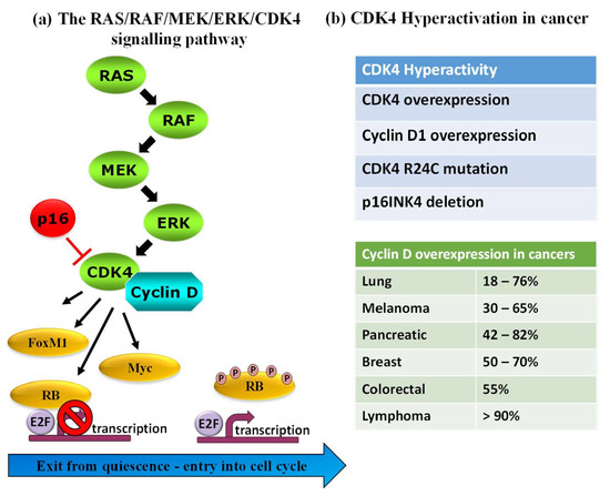
16 pages, 1333 KiB  
Review
Roles of AMPK and Its Downstream Signals in Pain Regulation
by Shenglan Wang and Yi Dai
Life 2021, 11(8), 836; https://doi.org/10.3390/life11080836 - 16 Aug 2021
Cited by 11 | Viewed by 4070
Abstract
Pain is an unpleasant sensory and emotional state that decreases quality of life. A metabolic sensor, adenosine monophosphate-activated protein kinase (AMPK), which is ubiquitously expressed in mammalian cells, has recently attracted interest as a new target of pain research. Abnormal AMPK expression and [...] Read more.
Pain is an unpleasant sensory and emotional state that decreases quality of life. A metabolic sensor, adenosine monophosphate-activated protein kinase (AMPK), which is ubiquitously expressed in mammalian cells, has recently attracted interest as a new target of pain research. Abnormal AMPK expression and function in the peripheral and central nervous systems are associated with various types of pain. AMPK and its downstream kinases participate in the regulation of neuron excitability, neuroinflammation and axonal and myelin regeneration. Numerous AMPK activators have reduced pain behavior in animal models. The current understanding of pain has been deepened by AMPK research, but certain issues, such as the interactions of AMPK at each step of pain regulation, await further investigation. This review examines the roles of AMPK and its downstream kinases in neurons and non-neuronal cells, as well as their contribution to pain regulation. Full article
Show Figures

Figure 1

Figure 1
<p>AMPK subunits and their activators in pain regulation. AICAR, 5-aminoimidazole-4-carboxamide ribonucleotide. ALA, α-lipoic acid. AMPK, adenosine monophosphate-activated protein kinase. CaMKKβ, calmodulin-dependent protein kinase kinase β. LKB1, liver kinase B1.</p>
Full article ">Figure 2
<p>Neuronal excitability is regulated by the AMPK/Nedd4-2 pathway. AMPK activation inhibits the binding of 14-3-3 with Nedd4-2 to increase Nedd4-2-mediated ubiquitination, which results in ion channel (e.g., TRPA1, Na<sub>v</sub>1.7, Na<sub>v</sub>1.8, Kv2.1) degradation in nociceptors. These events can subsequently suppress neuronal excitability. AMPK; adenosine monophosphate-activated protein kinase. DRG; dorsal root ganglion. Nedd4-2; neural precursor cell expressed developmentally downregulated 4-like kinase.</p>
Full article ">Figure 3
<p>The role of AMPK on neuroinflammation in microglial cells. AMPK induces autophagy by the direct phosphorylation of ULK1 or VPS34 and the indirect activation of ULK1 via inhibiting the mTORC1 pathway. Microglia can be stimulated to an M1 phenotype to release proinflammatory factors by defective autophagy induced by AMPK in pathological condition, while AMPK activator promotes microglial polarization to the M2 phenotype to release anti-inflammatory factors. AMPK; adenosine monophosphate-activated protein kinase. CSF1; colony stimulating factor 1. CSF1R; colony stimulating factor 1 receptor. DRG; dorsal root ganglion. LC3-II; microtubule-associated protein 1A/1B-light chain 3 (autophagosome membranes-associated form). LPS, lipopolysaccharides. mLST8, mammalian lethal with Sec13 protein 8. mTORC1, mammalian target of rapamycin complex 1. RAPTOR, regulatory protein associated with mTOR. TSC2, tuberous sclerosis complex 2. ULK1, unc-51 like autophagy activating kinase 1. VPS34, vacuolar protein sorting 34.</p>
Full article ">Figure 4
<p>Nerve regeneration is negatively regulated by AMPK. (<b>A</b>) The mTORC1 pathway inhibits cap-dependent mRNA degradation to increase transcription. Injury induces calcium influx to activate CaMKIIα/PSMC5 (26S proteasome subunit) pathway and degrades AMPK, resulting in mTORC1 pathway activation in axons. Decreased AMPK activation reduces cap-dependent mRNA degradation that is involved in axon regeneration. (<b>B</b>) Nerve injury impairs AMPK activation resulting in the upregulation of mTORC1 and c-jun pathways to enhance translation in Schwann cells. (<b>C</b>) AMPK activation inhibits axonal regeneration and Schwann cells-mediated re-myelination. AMPK, adenosine monophosphate-activated protein kinase. CaMKIIα, Ca<sup>2+</sup>/calmodulin-dependent protein kinase II α. elF4E, eukaryotic translation initiation factor. mTORC1, mammalian target of rapamycin complex 1. p4EBP, phosphorylated eukaryotic initiation factor-4E-binding protein. RAPTOR, regulatory protein associated with mTOR. TSC2, tuberous sclerosis complex 2.</p>
Full article ">
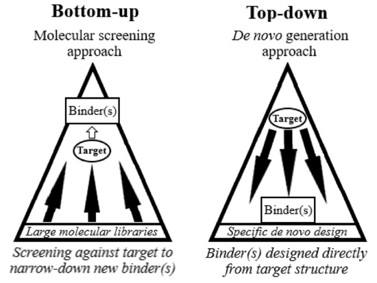
11 pages, 992 KiB  
Hypothesis
Evaluating the Microbial Habitability of Rogue Planets and Proposing Speculative Scenarios on How They Might Act as Vectors for Panspermia
by Dirk Schulze-Makuch and Alberto G. Fairén
Life 2021, 11(8), 833; https://doi.org/10.3390/life11080833 - 14 Aug 2021
Cited by 3 | Viewed by 4590
Abstract
There are two types of rogue planets, sub-brown dwarfs and “rocky” rogue planets. Sub-brown dwarfs are unlikely to be habitable or even host life, but rocky rogue planets may have a liquid ocean under a thick atmosphere or an ice layer. If they [...] Read more.
There are two types of rogue planets, sub-brown dwarfs and “rocky” rogue planets. Sub-brown dwarfs are unlikely to be habitable or even host life, but rocky rogue planets may have a liquid ocean under a thick atmosphere or an ice layer. If they are overlain by an insulating ice layer, they are also referred to as Steppenwolf planets. However, given the poor detectability of rocky rogue planets, there is still no direct evidence of the presence of water or ice on them. Here we discuss the possibility that these types of rogue planets could harbor unicellular organisms, conceivably based on a variety of different energy sources, including chemical, osmotic, thermal, and luminous energy. Further, given the theoretically predicted high number of rogue planets in the galaxy, we speculate that rogue planets could serve as a source for galactic panspermia, transferring life to other planetary systems. Full article
Show Figures

Figure 1

Figure 1
<p>Amount of ice necessary to create a liquid ocean on a Steppenwolf planet (modified from [<a href="#B20-life-11-00833" class="html-bibr">20</a>]).</p>
Full article ">Figure 2
<p>A sketch of how various types of thermotrophs could gain energy near a hydrothermal vent structure. In gray are marked the hotter areas within the model organism from which thermal energy can be obtained. In model (1), the thermotroph moves off the vent structure, but the vacuole is hotter and the putative organism can obtain energy from the thermal gradient. The principle is the same for (2) to (4), but the model organisms remain attached to the substrate. The heated vacuole moves within the cell (2), colder circulating water is heated within the organism (3), or an appendix of the cell moves in the thermal gradient back and forth (4) (modified from [<a href="#B40-life-11-00833" class="html-bibr">40</a>]).</p>
Full article ">
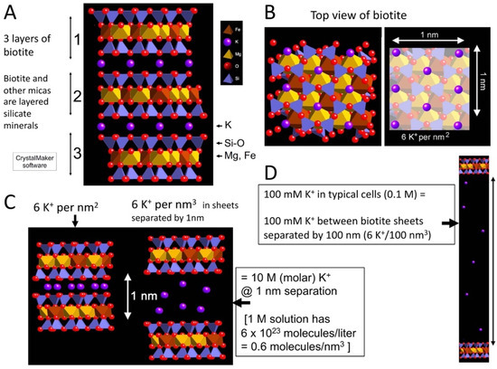
26 pages, 1599 KiB  
Review
Experimental Approaches for Testing the Hypothesis of the Emergence of Life at Submarine Alkaline Vents
by Thiago Altair, Luiz G. F. Borges, Douglas Galante and Hamilton Varela
Life 2021, 11(8), 777; https://doi.org/10.3390/life11080777 - 31 Jul 2021
Cited by 9 | Viewed by 4936
Abstract
Since the pioneering experimental work performed by Urey and Miller around 70 years ago, several experimental works have been developed for approaching the question of the origin of life based on very few well-constructed hypotheses. In recent years, attention has been drawn to [...] Read more.
Since the pioneering experimental work performed by Urey and Miller around 70 years ago, several experimental works have been developed for approaching the question of the origin of life based on very few well-constructed hypotheses. In recent years, attention has been drawn to the so-called alkaline hydrothermal vents model (AHV model) for the emergence of life. Since the first works, perspectives from complexity sciences, bioenergetics and thermodynamics have been incorporated into the model. Consequently, a high number of experimental works from the model using several tools have been developed. In this review, we present the key concepts that provide a background for the AHV model and then analyze the experimental approaches that were motivated by it. Experimental tools based on hydrothermal reactors, microfluidics and chemical gardens were used for simulating the environments of early AHVs on the Hadean Earth (~4.0 Ga). In addition, it is noteworthy that several works used techniques from electrochemistry to investigate phenomena in the vent–ocean interface for early AHVs. Their results provided important parameters and details that are used for the evaluation of the plausibility of the AHV model, and for the enhancement of it. Full article
Show Figures

Figure 1

Figure 1
<p>Comparison between the chemiosmotic mechanism on modern cells and the proposed mechanism for the vent-ocean interface: (<b>a</b>) typical setting for the chemiosmotic mechanism synthesizing ATP; (<b>b</b>) electron and proton transport coupled proposed for CO<sub>2</sub> reduction on a membrane [<a href="#B92-life-11-00777" class="html-bibr">92</a>]; (<b>c</b>) proposed prebiotic chemiosmotic mechanism for CO<sub>2</sub> reduction in a Hadean vent–ocean interface.</p>
Full article ">Figure 2
<p>Visual illustration of the chemical reactors simulating early vent-ocean environment: (<b>a</b>) the reactor column from the hydrothermal setup developed by Mielke et al., 2010 [<a href="#B105-life-11-00777" class="html-bibr">105</a>] and adapted by White et al., 2020 [<a href="#B53-life-11-00777" class="html-bibr">53</a>] for simulating the contact of hydrothermal fluid, early ocean fluid and the Hadean crust under high pressure; (<b>b</b>) the origin-of-life reactor, as proposed by Herschy et al. (2014) [<a href="#B106-life-11-00777" class="html-bibr">106</a>] main vessel for testing the formation of Fe(Ni)S barrier and its properties for organic formation.</p>
Full article ">Figure 3
<p>The analogy between iron–sulfur chemical gardens in a laboratory experiment (<b>a</b>) and an AHV mound on the Hadean ocean floor (<b>b</b>) regarding the generated natural membrane potential (∆E) as a consequence of pH gradient (∆pH) and redox gradient (∆ϕ). In addition, (<b>c</b>) a typically simplified scheme for the protocol that measures the membrane potential results in the values shown in <a href="#life-11-00777-t001" class="html-table">Table 1</a>.</p>
Full article ">
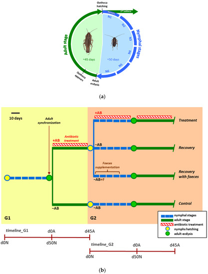
13 pages, 544 KiB  
Review
Role of Matrix Gla Protein in the Complex Network of Coronary Artery Disease: A Comprehensive Review
by Marko Kumric, Josip A. Borovac, Tina Ticinovic Kurir, Dinko Martinovic, Ivan Frka Separovic, Ljupka Baric and Josko Bozic
Life 2021, 11(8), 737; https://doi.org/10.3390/life11080737 - 24 Jul 2021
Cited by 7 | Viewed by 3378
Abstract
Coronary artery disease (CAD) is widely recognized as one of the most important clinical entities. In recent years, a large body of accumulated data suggest that coronary artery calcification, a process highly prevalent in patients with CAD, occurs via well-organized biologic processes, rather [...] Read more.
Coronary artery disease (CAD) is widely recognized as one of the most important clinical entities. In recent years, a large body of accumulated data suggest that coronary artery calcification, a process highly prevalent in patients with CAD, occurs via well-organized biologic processes, rather than passively, as previously regarded. Matrix Gla protein (MGP), a vitamin K-dependent protein, emerged as an important inhibitor of both intimal and medial vascular calcification. The functionality of MGP hinges on two post-translational modifications: phosphorylation and carboxylation. Depending on the above-noted modifications, various species of MGP may exist in circulation, each with their respective level of functionality. Emerging data suggest that dysfunctional species of MGP, markedly, dephosphorylated-uncarboxylated MGP, might find its application as biomarkers of microvascular health, and assist in clinical decision making with regard to initiation of vitamin K supplementation. Hence, in this review we summarized the current knowledge with respect to the role of MGP in the complex network of vascular calcification with concurrent inferences to CAD. In addition, we discussed the effects of warfarin use on MGP functionality, with concomitant implications to coronary plaque stability. Full article
Show Figures

Figure 1

Figure 1
<p>Post-translational modifications of MGP and the mechanisms by which MGP is implicated in vascular calcification. Red lines depict inhibition of the process, whereas the blue line indicate stimulation.</p>
Full article ">
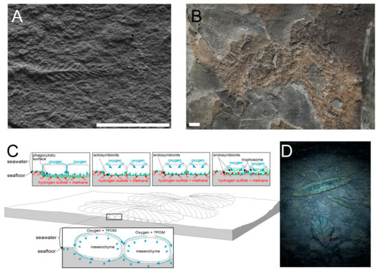
16 pages, 633 KiB  
Review
Non-Coding RNAs and Splicing Activity in Testicular Germ Cell Tumors
by Marco Barchi, Pamela Bielli, Susanna Dolci, Pellegrino Rossi and Paola Grimaldi
Life 2021, 11(8), 736; https://doi.org/10.3390/life11080736 - 24 Jul 2021
Cited by 9 | Viewed by 3868
Abstract
Testicular germ cell tumors (TGCTs) are the most common tumors in adolescent and young men. Recently, genome-wide studies have made it possible to progress in understanding the molecular mechanisms underlying the development of tumors. It is becoming increasingly clear that aberrant regulation of [...] Read more.
Testicular germ cell tumors (TGCTs) are the most common tumors in adolescent and young men. Recently, genome-wide studies have made it possible to progress in understanding the molecular mechanisms underlying the development of tumors. It is becoming increasingly clear that aberrant regulation of RNA metabolism can drive tumorigenesis and influence chemotherapeutic response. Notably, the expression of non-coding RNAs as well as specific splice variants is deeply deregulated in human cancers. Since these cancer-related RNA species are considered promising diagnostic, prognostic and therapeutic targets, understanding their function in cancer development is becoming a major challenge. Here, we summarize how the different expression of RNA species repertoire, including non-coding RNAs and protein-coding splicing variants, impacts on TGCTs’ onset and progression and sustains therapeutic resistance. Finally, the role of transcription-associated R-loop misregulation in the maintenance of genomic stability in TGCTs is also discussed. Full article
Show Figures

Figure 1

Figure 1
<p>Schematic representation of non-coding RNAs and their biological functions.</p>
Full article ">
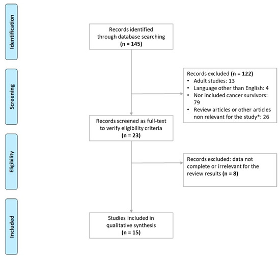
12 pages, 2845 KiB  
Review
Transforaminal Lumbar Interbody Fusion (TLIF) versus Oblique Lumbar Interbody Fusion (OLIF) in Interbody Fusion Technique for Degenerative Spondylolisthesis: A Systematic Review and Meta-Analysis
by Min Cheol Chang, Gang-Un Kim, Yoo Jin Choo and Gun Woo Lee
Life 2021, 11(7), 696; https://doi.org/10.3390/life11070696 - 15 Jul 2021
Cited by 23 | Viewed by 7235
Abstract
Preoperative pathology requiring fusion surgery has a great impact on postoperative outcomes. However, the previous clinical and meta-analysis studies did not control for the pathology. In this systematic review, the authors aimed to compare oblique lumbar interbody fusion (OLIF) with transforaminal interbody fusion [...] Read more.
Preoperative pathology requiring fusion surgery has a great impact on postoperative outcomes. However, the previous clinical and meta-analysis studies did not control for the pathology. In this systematic review, the authors aimed to compare oblique lumbar interbody fusion (OLIF) with transforaminal interbody fusion (TLIF) as an interbody fusion technique in lumbar fusion surgery for patients with degenerative spondylolisthesis (DS). We systematically searched for relevant articles in the available databases. Among the 3022 articles, three studies were identified and met the inclusion criteria. In terms of radiological outcome, the amount of disc height restoration was greater in the OLIF group than in the TLIF group, but there was no significant difference between the two surgical techniques (p = 0.18). In the clinical outcomes, the pain improvement was not significantly different between the two surgical techniques. In terms of surgical outcomes, OLIF resulted in a shorter length of hospital stay and less blood loss than TLIF (p < 0.0001 and p = 0.02, respectively). The present meta-analysis indicated no significant difference in clinical, radiological outcomes, and surgical time between TLIF and OLIF for DS, but the lengths of hospital stay and blood loss were better in OLIF than TLIF. Though encouraging, these findings were based on low-quality evidence from a small number of retrospective studies that are prone to bias. Full article
Show Figures

Figure 1

Figure 1
<p>Flow chart showing the search results of the meta-analysis.</p>
Full article ">Figure 2
<p>Results of meta-analysis for treatment outcome.</p>
Full article ">Figure 3
<p>Results of meta-analysis for surgical outcome.</p>
Full article ">Figure 4
<p>Graphic funnel plots of the included studies.</p>
Full article ">Figure 5
<p>Illustration about lumbar interbody fusion techniques.</p>
Full article ">
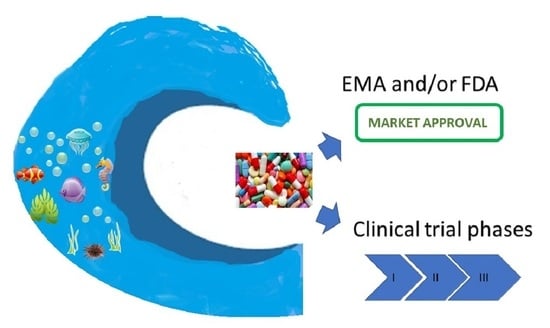
26 pages, 2065 KiB  
Review
‘Whole Organism’, Systems Biology, and Top-Down Criteria for Evaluating Scenarios for the Origin of Life
by Clifford F. Brunk and Charles R. Marshall
Life 2021, 11(7), 690; https://doi.org/10.3390/life11070690 - 14 Jul 2021
Cited by 16 | Viewed by 6250
Abstract
While most advances in the study of the origin of life on Earth (OoLoE) are piecemeal, tested against the laws of chemistry and physics, ultimately the goal is to develop an overall scenario for life’s origin(s). However, the dimensionality of non-equilibrium chemical systems, [...] Read more.
While most advances in the study of the origin of life on Earth (OoLoE) are piecemeal, tested against the laws of chemistry and physics, ultimately the goal is to develop an overall scenario for life’s origin(s). However, the dimensionality of non-equilibrium chemical systems, from the range of possible boundary conditions and chemical interactions, renders the application of chemical and physical laws difficult. Here we outline a set of simple criteria for evaluating OoLoE scenarios. These include the need for containment, steady energy and material flows, and structured spatial heterogeneity from the outset. The Principle of Continuity, the fact that all life today was derived from first life, suggests favoring scenarios with fewer non-analog (not seen in life today) to analog (seen in life today) transitions in the inferred first biochemical pathways. Top-down data also indicate that a complex metabolism predated ribozymes and enzymes, and that full cellular autonomy and motility occurred post-LUCA. Using these criteria, we find the alkaline hydrothermal vent microchamber complex scenario with a late evolving exploitation of the natural occurring pH (or Na+ gradient) by ATP synthase the most compelling. However, there are as yet so many unknowns, we also advocate for the continued development of as many plausible scenarios as possible. Full article
Show Figures

Figure 1

Figure 1
<p>Terminology used in this paper. ‘Crown Group Life’ refers to the most recent common ancestor of all living things (LUCA) and all its descendants (regardless of whether they are extinct or not). ‘Stem Group Life’ refers to all living things (now extinct) that predate LUCA, including the Initial Darwinian Ancestor (IDA) [<a href="#B37-life-11-00690" class="html-bibr">37</a>] back to the hard-to-define transition between prebiotic chemistry and Pre-Darwinian Life, which consists of systems that might be viewed as being alive, but not yet capable of reproduction. The terms crown and stem can be applied to any lineage, as shown here for the Archaea. The grey inset is meant to convey the likelihood that there was extensive exchange of information and molecules between entities that might be viewed as being alive throughout all the early phases of life’s history. Note that we have not included the eukaryotes in this figure.</p>
Full article ">Figure 2
<p>Structure of the (<b>a</b>) terrestrial wet–dry cycles [<a href="#B21-life-11-00690" class="html-bibr">21</a>] and (<b>b</b>) marine microchamber complex scenarios for the origin of life [<a href="#B12-life-11-00690" class="html-bibr">12</a>]. Terrestrial wet–dry cycles scenarios typically posit an energy source (UV light) and source materials (HCN) not used by life today, and thus are designated non-analog. Similarly, the microchamber complex scenario posits that initial containment was provided by the microchamber walls, not used by life today, and so is also designated as non-analog. The dashed lines indicate steps that are not (currently) well explained by the scenario.</p>
Full article ">Figure 3
<p>Conceptual histogram expressing our sense that the pre-Darwinian stage of life emerged a great many times, while relatively few made the difficult transition to the “plus-RNA-World”, with even less achieving protein synthesis, followed by perhaps an easier transition to emergence from the microchambers as free-living cells. Reflecting our ignorance, we have assigned frequencies in terms of 10<span class="html-italic"><sup>x</sup></span>, where the value of <span class="html-italic">x</span> is unknown. If <span class="html-italic">x</span> = 12, then the scaling of the histogram indicates 1,000,000,000,000 origins of prebiotic metabolism, 1000 “plus-RNA-Worlds”, 10 origins of protein synthesis, and six emergences, which implies the extinction of four of them.</p>
Full article ">Figure 4
<p>Hypothesized sequence of major events in the origin and early evolution of life within the ancestral microchamber complex, with a focus on the pre-Darwinian stages, and on the emergence of the Bacteria and Archaea. Innovations in red are emphasized in the text. Most of the events depicted occurred within the confines of the vent microchambers; only at the very top do Bacteria and Archaea emerge as free-living cells. The developments prior to IDA are relatively slow due to limited energy and nonspecific catalysts. Beginning with IDA, Darwinian evolution accelerated the rate of change, while between IDA and LUCA ribozymes were replaced by enzymes. Eventually energy from the natural proton gradient, harnessed by ATP synthase, provided an energy source in addition to the H<sub>2</sub> from serpentinization. Following LUCA, the progenitors of Bacteria and Archaea evolved within the microchamber complex prior to their emergence as free-living cells. Life as defined here includes both stem and crown group life (see <a href="#life-11-00690-f001" class="html-fig">Figure 1</a>). Note we have not depicted the likely extensive role of horizontal transfer in these early phases of life’s history (but see <a href="#life-11-00690-f001" class="html-fig">Figure 1</a>).</p>
Full article ">
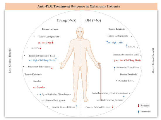
16 pages, 1073 KiB  
Review
Hyperalphalipoproteinemia and Beyond: The Role of HDL in Cardiovascular Diseases
by Antonina Giammanco, Davide Noto, Carlo Maria Barbagallo, Emilio Nardi, Rosalia Caldarella, Marcello Ciaccio, Maurizio Rocco Averna and Angelo Baldassare Cefalù
Life 2021, 11(6), 581; https://doi.org/10.3390/life11060581 - 18 Jun 2021
Cited by 12 | Viewed by 4947
Abstract
Hyperalphalipoproteinemia (HALP) is a lipid disorder characterized by elevated plasma high-density lipoprotein cholesterol (HDL-C) levels above the 90th percentile of the distribution of HDL-C values in the general population. Secondary non-genetic factors such as drugs, pregnancy, alcohol intake, and liver diseases might induce [...] Read more.
Hyperalphalipoproteinemia (HALP) is a lipid disorder characterized by elevated plasma high-density lipoprotein cholesterol (HDL-C) levels above the 90th percentile of the distribution of HDL-C values in the general population. Secondary non-genetic factors such as drugs, pregnancy, alcohol intake, and liver diseases might induce HDL increases. Primary forms of HALP are caused by mutations in the genes coding for cholesteryl ester transfer protein (CETP), hepatic lipase (HL), apolipoprotein C-III (apo C-III), scavenger receptor class B type I (SR-BI) and endothelial lipase (EL). However, in the last decades, genome-wide association studies (GWAS) have also suggested a polygenic inheritance of hyperalphalipoproteinemia. Epidemiological studies have suggested that HDL-C is inversely correlated with cardiovascular (CV) risk, but recent Mendelian randomization data have shown a lack of atheroprotective causal effects of HDL-C. This review will focus on primary forms of HALP, the role of polygenic inheritance on HDL-C, associated risk for cardiovascular diseases and possible treatment options. Full article
Show Figures

Figure 1

Figure 1
<p>Schematic representation of the biogenesis of HDL. The first step in HDL biogenesis begins with the secretion of Apo-A1 by the liver and the intestine. The lipid-poor Apo-A1 then interacts with ABCA1 and progressively gains phospholipids (PL) and free cholesterol (FC) from the cells. The lipidated apoA-I is gradually converted to discoidal particles composed of unesterified cholesterol. Then, the enzyme lecithin/cholesterol acyltransferase (LCAT) esterifies the FC and the discoidal HDLs are finally converted to spherical HDL particles containing Apo-A1, Apo-E or Apo-A4: secondary causes of hyperalphalipoproteinemia.</p>
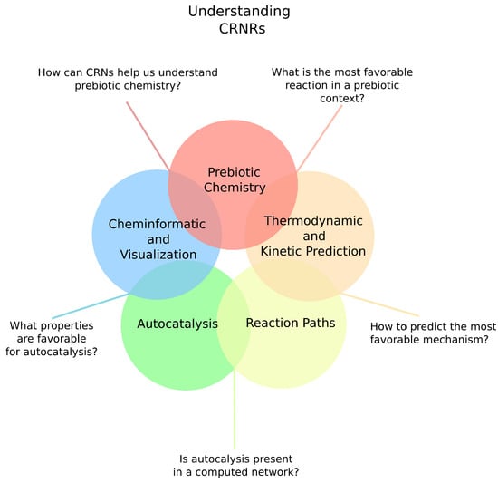
Full article ">Figure 2
<p>Schematic representation of the Reverse Cholesterol Transport (RCT) mechanism. In the RCT process, the ApoA-I receives the cholesterol from the foam cells (macrophages) through the ATP-binding cassette transporter member 1 (ABCA1) (<b>1</b>), leading HDL to become mature in a mechanism mediated by the transporter ABCG1 (<b>2</b>). The Lecitin:cholesterol acyltransferase (LCAT) (<b>3</b>) esterifies the free cholesterol (FC) thus contributing to form mature and spherical HDLs formed of a cholesteryl ester (CE) core. Finally, HDLs bind to the scavenger receptor class B type-1 (SR-B1) in the liver (<b>4</b>) and are selectively removed from the blood stream.</p>
Full article ">
17 pages, 888 KiB  
Review
Molecular Mechanisms of Action of Novel Psychoactive Substances (NPS). A New Threat for Young Drug Users with Forensic-Toxicological Implications
by Arianna Giorgetti, Jennifer P. Pascali, Paolo Fais, Guido Pelletti, Andrea Gabbin, Giorgia Franchetti, Giovanni Cecchetto and Guido Viel
Life 2021, 11(5), 440; https://doi.org/10.3390/life11050440 - 14 May 2021
Cited by 17 | Viewed by 4952
Abstract
Novel psychoactive substances (NPS) represent a severe health risk for drug users. Even though the phenomenon has been growing since the early 2000s, the mechanisms of action of NPS at the receptors and beyond them are still scarcely understood. The aim of the [...] Read more.
Novel psychoactive substances (NPS) represent a severe health risk for drug users. Even though the phenomenon has been growing since the early 2000s, the mechanisms of action of NPS at the receptors and beyond them are still scarcely understood. The aim of the present study was to provide a systematic review of the updated knowledge regarding the molecular mechanisms underlying the toxicity of synthetic opioids, cannabinoids, cathinones, and stimulants. The study was conducted on the PubMed database. Study eligibility criteria included relevance to the topic, English language, and time of publication (2010–2020). A combined Mesh and free-text protocols search was performed. Study selection was performed on the title/abstract and, in doubtful cases, on the full texts of papers. Of the 580 records identified through PubMed searching and reference checking, 307 were excluded by title/abstract and 78 additional papers were excluded after full-text reading, leaving a total of 155 included papers. Molecular mechanisms of synthetic opioids, synthetic cannabinoids, stimulants, psychedelics, and hallucinogens were reviewed and mostly involved both a receptor-mediated and non-receptor mediated cellular modulation with multiple neurotransmitters interactions. The molecular mechanisms underlying the action of NPS are more complex than expected, with a wide range of overlap among activated receptors and neurotransmitter systems. The peculiar action profile of single compounds does not necessarily reflect that of the structural class to which they belong, accounting for possible unexpected toxic reactions. Full article
Show Figures

Figure 1

Figure 1
<p>PRISMA flow diagram of the present review. From: Moher D, Liberati A, Tetzlaff J, et al. Preferred reporting items for systematic reviews and meta-analyses: the PRISMA statement. PLoS medicine, 2009, 6(7): e1000097 [<a href="#B8-life-11-00440" class="html-bibr">8</a>].</p>
Full article ">Figure 2
<p>Modified from Guzman et al. Cannabinoids: Potential anticancer agents. <span class="html-italic">Nat Rev Cancer</span>. Mechanism activated by the receptor of human cannabinoids 1 (CB1R), ranging from binding to G-protein-coupled receptors (Gi/o) with inhibition of the adenylyl cyclase (AdeC), and therefore of the cyclicAMP (cAMP) and of the protein kinase A (PKA). Inhibition of voltage-sensitive Ca<sup>2+</sup> channels (VSCC); release of Ca<sup>2+</sup> from intracellular stores; activation of the phosphatidylinositol 3-kinase (PI3K)–AKT pathway; activation of mitogen-activated protein kinase cascades as extracellular-signal-regulated kinase (ERK), JUN amino-terminal kinase (JNK), and p38 and ceramide generation through FAN–sphingomyelinase (factor associated with neutral sphingomyelinase activation–SMase).</p>
Full article ">Figure 3
<p>Selectivity of stimulants with the ratio between dopamine (DAT) and serotonin (SERT) transporters. Slightly modified from Luethi et al. [<a href="#B7-life-11-00440" class="html-bibr">7</a>].</p>
Full article ">
28 pages, 3121 KiB  
Review
The “Water Problem”(sic), the Illusory Pond and Life’s Submarine Emergence—A Review
by Michael J. Russell
Life 2021, 11(5), 429; https://doi.org/10.3390/life11050429 - 10 May 2021
Cited by 25 | Viewed by 8049
Abstract
The assumption that there was a “water problem” at the emergence of life—that the Hadean Ocean was simply too wet and salty for life to have emerged in it—is here subjected to geological and experimental reality checks. The “warm little pond” that would [...] Read more.
The assumption that there was a “water problem” at the emergence of life—that the Hadean Ocean was simply too wet and salty for life to have emerged in it—is here subjected to geological and experimental reality checks. The “warm little pond” that would take the place of the submarine alkaline vent theory (AVT), as recently extolled in the journal Nature, flies in the face of decades of geological, microbiological and evolutionary research and reasoning. To the present author, the evidence refuting the warm little pond scheme is overwhelming given the facts that (i) the early Earth was a water world, (ii) its all-enveloping ocean was never less than 4 km deep, (iii) there were no figurative “Icelands” or “Hawaiis”, nor even an “Ontong Java” then because (iv) the solidifying magma ocean beneath was still too mushy to support such salient loadings on the oceanic crust. In place of the supposed warm little pond, we offer a well-protected mineral mound precipitated at a submarine alkaline vent as life’s womb: in place of lipid membranes, we suggest peptides; we replace poisonous cyanide with ammonium and hydrazine; instead of deleterious radiation we have the appropriate life-giving redox and pH disequilibria; and in place of messy chemistry we offer the potential for life’s emergence from the simplest of geochemically available molecules and ions focused at a submarine alkaline vent in the Hadean—specifically within the nano-confined flexible and redox active interlayer walls of the mixed-valent double layer oxyhydroxide mineral, fougerite/green rust comprising much of that mound. Full article
Show Figures

Figure 1

Figure 1
<p>Depiction of our Hadean planet. The crust was completely submerged with a ~5 km deep ocean as the magma ocean was still too mushy to support significant bulges even at the apices of mantle plumes [<a href="#B98-life-11-00429" class="html-bibr">98</a>,<a href="#B99-life-11-00429" class="html-bibr">99</a>,<a href="#B100-life-11-00429" class="html-bibr">100</a>,<a href="#B101-life-11-00429" class="html-bibr">101</a>,<a href="#B102-life-11-00429" class="html-bibr">102</a>,<a href="#B106-life-11-00429" class="html-bibr">106</a>,<a href="#B233-life-11-00429" class="html-bibr">233</a>,<a href="#B234-life-11-00429" class="html-bibr">234</a>,<a href="#B235-life-11-00429" class="html-bibr">235</a>] EoL: emergence of life.</p>
Full article ">
15 pages, 1087 KiB  
Review
Chronothyroidology: Chronobiological Aspects in Thyroid Function and Diseases
by Giuseppe Bellastella, Maria Ida Maiorino, Lorenzo Scappaticcio, Annamaria De Bellis, Silvia Mercadante, Katherine Esposito and Antonio Bellastella
Life 2021, 11(5), 426; https://doi.org/10.3390/life11050426 - 10 May 2021
Cited by 11 | Viewed by 3788
Abstract
Chronobiology is the scientific discipline which considers biological phenomena in relation to time, which assumes itself biological identity. Many physiological processes are cyclically regulated by intrinsic clocks and many pathological events show a circadian time-related occurrence. Even the pituitary–thyroid axis is under the [...] Read more.
Chronobiology is the scientific discipline which considers biological phenomena in relation to time, which assumes itself biological identity. Many physiological processes are cyclically regulated by intrinsic clocks and many pathological events show a circadian time-related occurrence. Even the pituitary–thyroid axis is under the control of a central clock, and the hormones of the pituitary–thyroid axis exhibit circadian, ultradian and circannual rhythmicity. This review, after describing briefly the essential principles of chronobiology, will be focused on the results of personal experiences and of other studies on this issue, paying particular attention to those regarding the thyroid implications, appearing in the literature as reviews, metanalyses, original and observational studies until 28 February 2021 and acquired from two databases (Scopus and PubMed). The first input to biological rhythms is given by a central clock located in the suprachiasmatic nucleus (SCN), which dictates the timing from its hypothalamic site to satellite clocks that contribute in a hierarchical way to regulate the physiological rhythmicity. Disruption of the rhythmic organization can favor the onset of important disorders, including thyroid diseases. Several studies on the interrelationship between thyroid function and circadian rhythmicity demonstrated that thyroid dysfunctions may affect negatively circadian organization, disrupting TSH rhythm. Conversely, alterations of clock machinery may cause important perturbations at the cellular level, which may favor thyroid dysfunctions and also cancer. Full article
Show Figures

Figure 1

Figure 1
<p>Schematic representation of a circadian virtual rhythm and its parameters (period, zenith or acrophase, mean level or mesor, amplitude), obtained by fitting a sinusoidal curve to a naïf pattern of data by the “least squares” method: Period of 24 h = 360° ([<a href="#B13-life-11-00426" class="html-bibr">13</a>], reproduced with permission of the Editor).</p>
Full article ">Figure 2
<p>Synchronization by light input of the rhythmic variations in hypothalamic–pituitary–thyroid secretions: It acts directly, through the retino-hypothalmic tract (RHT), by exciting the light-entrainable circadian pacemaker located in the suprachiasmatic nucleus (SCN) of the hypothalamus, which then outputs the circadian signal via neural projection, exciting the rhythmic secretion of thyrotropin-releasing hormone (TRH) and consequently of thyrotropin (TSH) and thyroid hormones(T4:thyroxine; T3:triiodotyronine). Light acts also indirectly, by modulating with an inhibiting effect the variations in melatonin secretion at retina and pineal gland levels, thus further stimulating the secretions of the hypothalamic–pituitary–thyroid axis.</p>
Full article ">Figure 3
<p>Schematic representation of physiological organization of rhythmic hypothalamic–pituitary–thyroid secretions: TRH(thyrotropin-releasing hormone) and somatostatin from the hypothalamus exert a stimulating and inhibiting action, respectively, on the pituitary secretion of TSH(thyrotropin), which in turn stimulates the thyroid gland to produce T4(thyroxine) and T3(triiodotyronine). Thyroid hormones regulate, by a negative feedback, the secretion of TRH and TSH, by acting both at hypothalamus and pituitary level.</p>
Full article ">Figure 4
<p>Circadian and ultradian variations in plasma TSH (thyrotropin)concentrations obtained by frequent samples over 24 h in a single healthy adult volunteer. The black bar indicates the period of sleep, the arrows the time of the meals.</p>
Full article ">Figure 5
<p>(<b>A</b>) Monthly mean (±SD) of TSH, T4, T3, GH values in prepubertal subjects. (<b>B</b>) Single cosinor display of circannual variations in these hormone concentrations and their characteristics, visualized by polar cosinor plot. A significant rhythm was validated only for TSH, as revealed by its ellipse (continuous tract), which does not cover the pole (zero: center), and with acrophase in December, as indicated by the arrow starting from the center and directed towards December (confidence limits October–February) [<a href="#B68-life-11-00426" class="html-bibr">68</a>].</p>
Full article ">
9 pages, 234 KiB  
Review
Different Chronic Disorders That Fall within the Term Juvenile Idiopathic Arthritis
by Lucia M. Sur, Remus Gaga, Emanuela Duca, Genel Sur, Iulia Lupan, Daniel Sur, Gabriel Samasca, Cecilia Lazea and Calin Lazar
Life 2021, 11(5), 398; https://doi.org/10.3390/life11050398 - 27 Apr 2021
Cited by 6 | Viewed by 2534
Abstract
Juvenile idiopathic arthritis (JIA) represents a significant challenge for pediatricians who intend to diagnose and treat this pathology. The classification criteria for JIA subtypes are rigid and often do not fully satisfy the possibilities of classification in the subtype. The objective of this [...] Read more.
Juvenile idiopathic arthritis (JIA) represents a significant challenge for pediatricians who intend to diagnose and treat this pathology. The classification criteria for JIA subtypes are rigid and often do not fully satisfy the possibilities of classification in the subtype. The objective of this study was to identify clearer criteria for classifying JIA subtypes. The 2019 expert committee meeting (PRINTO) shows the difficulties of this classification and proposes new research directions for the identification of disease subtypes. Four different chronic disorders are used to define JIA in a concise and easy to follow classification system. However, dates from the literature suggest that at least 10% of cases are still difficult to classify. Possibly in the future, different classifications of JIA based on pathophysiological and genetic criteria would be necessary. Full article
20 pages, 7513 KiB  
Review
The “Genomic Code”: DNA Pervasively Moulds Chromatin Structures Leaving no Room for “Junk”
by Giorgio Bernardi
Life 2021, 11(4), 342; https://doi.org/10.3390/life11040342 - 13 Apr 2021
Cited by 8 | Viewed by 3720
Abstract
The chromatin of the human genome was analyzed at three DNA size levels. At the first, compartment level, two “gene spaces” were found many years ago: A GC-rich, gene-rich “genome core” and a GC-poor, gene-poor “genome desert”, the former corresponding to open chromatin [...] Read more.
The chromatin of the human genome was analyzed at three DNA size levels. At the first, compartment level, two “gene spaces” were found many years ago: A GC-rich, gene-rich “genome core” and a GC-poor, gene-poor “genome desert”, the former corresponding to open chromatin centrally located in the interphase nucleus, the latter to closed chromatin located peripherally. This bimodality was later confirmed and extended by the discoveries (1) of LADs, the Lamina-Associated Domains, and InterLADs; (2) of two “spatial compartments”, A and B, identified on the basis of chromatin interactions; and (3) of “forests and prairies” characterized by high and low CpG islands densities. Chromatin compartments were shown to be associated with the compositionally different, flat and single- or multi-peak DNA structures of the two, GC-poor and GC-rich, “super-families” of isochores. At the second, sub-compartment, level, chromatin corresponds to flat isochores and to isochore loops (due to compositional DNA gradients) that are susceptible to extrusion. Finally, at the short-sequence level, two sets of sequences, GC-poor and GC-rich, define two different nucleosome spacings, a short one and a long one. In conclusion, chromatin structures are moulded according to a “genomic code” by DNA sequences that pervade the genome and leave no room for “junk”. Full article
Show Figures

Figure 1

Figure 1
<p>(<b>A</b>) Isochore families. The histogram displays the isochores from the human genome as pooled in bins of 1% GC. The Gaussian profile shows the distribution of isochore families, which are represented in different colours. Gene densities define a “genome desert”, comprising isochore families L1 and L2<sup>−</sup>, and a “genome core”, comprising isochore families L2<sup>+</sup>, H1, H2, H3; L2<sup>−</sup> and L2<sup>+</sup> sub-families are separated by a vertical broken red line. (Modified from [<a href="#B37-life-11-00342" class="html-bibr">37</a>]). (<b>B</b>) Gene distribution in the isochore families of the bovine genome. (Drawn from data of [<a href="#B37-life-11-00342" class="html-bibr">37</a>]); Two different lines of gene concentration characterized by different slopes were drawn through L1-H1 and H1-H3 points. (<b>C</b>) A FISH pattern of H2/H3 and L1 isochores (a clearer version of Figure 2A3 of [<a href="#B38-life-11-00342" class="html-bibr">38</a>]) is compared with a Hi-C pattern (modified from [<a href="#B39-life-11-00342" class="html-bibr">39</a>]), in which case compartment A is central, compartment B is peripheral and nucleolar.</p>
Full article ">Figure 2
<p>(<b>A</b>) The inverted LAD profile of the long arm of human chromosome 21 (from [<a href="#B81-life-11-00342" class="html-bibr">81</a>]) is compared with the 100 Kb dot-plot GC profile of <a href="#life-11-00342-f003" class="html-fig">Figure 3</a>B (from [<a href="#B7-life-11-00342" class="html-bibr">7</a>]). LADs correspond to L1 isochores (two blue lines bracket region 2, the largest L1 isochore) and to two “valley” isochores (double black lines, X and Y, that belong in the L2<sup>-</sup> sub-family, a low heterogeneity subfamily in the L2 isochore GC range). One H1 (a), and five L2<sup>+</sup> single peak isochores (b–f) correspond to InterLADs (the L2<sup>+</sup> sub-family is a heterogeneous sub-family in the L2 isochore GC range). (<b>C</b>) The 100 Kb profile is compared with the A and B compartments (adapted from [<a href="#B82-life-11-00342" class="html-bibr">82</a>]; the thickness of the profile is due to the enlargement of the original minute figure). Imperfect alignments of Figures (<b>A</b>–<b>C</b>) are due to the different original sources of the panels. (<b>D</b>) The enlarged GC profiles of regions 2 and 6 (corresponding to the telomere) show the striking differences in their compositional ranges (see text).</p>
Full article ">Figure 3
<p>(<b>A</b>) A profile of a chromosome segment in which all isochore families are represented is compared with the corresponding compartment profile to show that, while the L1 and L2<sup>−</sup> isochores correspond to the B compartment, the other isochores correspond to the A compartments (modified from [<a href="#B6-life-11-00342" class="html-bibr">6</a>]). (<b>B</b>) The figure shows an average match between isochores/isochore-blocks (profiles) and chromatin sub-compartments (A1–B4). Blue/black triangles are examples of good/bad matches (from a Supplementary Figure of [<a href="#B30-life-11-00342" class="html-bibr">30</a>]).</p>
Full article ">Figure 4
<p>(<b>A</b>) ~30 Mb of human chromosome 7 is analyzed at a resolution of 50 Kb; a fine correspondence of isochore boundaries with LAD and TAD boundaries can be observed (modified from [<a href="#B30-life-11-00342" class="html-bibr">30</a>]). (<b>B</b>) The chromatin loops from a 2.1 Mb region of human chromosome 20 have been aligned with the corresponding heat map which was used to segment the corresponding DNA sequence into isochores; asterisks indicate small anomalies in the isochores/domains correspondence (modified from [<a href="#B30-life-11-00342" class="html-bibr">30</a>]).</p>
Full article ">Figure 5
<p>The proposed model for the formation of “primary TADs” and TADs. (Modified from [<a href="#B8-life-11-00342" class="html-bibr">8</a>]; See text).</p>
Full article ">Figure 6
<p>(<b>A</b>,<b>B</b>) Profiles of tri- and penta- A’s and G’s of regions 2 and 6 (from <a href="#app1-life-11-00342" class="html-app">Figure S3</a> of [<a href="#B108-life-11-00342" class="html-bibr">108</a>] (<b>C</b>) A scheme of nucleosome densities for regions 2 and 6. (Modified from [<a href="#B109-life-11-00342" class="html-bibr">109</a>]; See also legend of <a href="#life-11-00342-f007" class="html-fig">Figure 7</a>).</p>
Full article ">Figure 7
<p>Functional elements across the human genome [<a href="#B109-life-11-00342" class="html-bibr">109</a>]. This Figure can also be seen as a model representing the structural elements discussed in the article (see text). This figure is published with the permission of authors and Springer Nature.</p>
Full article ">
Back to TopTop