Category:Dental diagrams
Zur Navigation springen
Zur Suche springen
Zahnbezeichnungen des Menschen in verschiedenen Schemata | |||||
| Medium hochladen | |||||
| Unterklasse von | |||||
|---|---|---|---|---|---|
| Verwendung | |||||
| |||||
Unterkategorien
Es werden 3 von insgesamt 3 Unterkategorien in dieser Kategorie angezeigt:
In Klammern die Anzahl der enthaltenen Kategorien (K), Seiten (S), Dateien (D)
D
- Diagrams of sets of teeth (54 D)
P
Medien in der Kategorie „Dental diagrams“
Folgende 199 Dateien sind in dieser Kategorie, von 199 insgesamt.
-
110216ek08.jpg 1.024 × 717; 111 KB
-
202402 Oral Cavity.svg 512 × 512; 675 KB
-
202402 Periodontal Disease.svg 512 × 512; 438 KB
-
202402 Tooth Structure.svg 512 × 512; 327 KB
-
3 bild.jpg 241 × 282; 35 KB
-
4 bild.jpg 320 × 253; 35 KB
-
4b bild.jpg 550 × 327; 65 KB
-
5 bild.jpg 280 × 327; 45 KB
-
5b bild.jpg 433 × 195; 40 KB
-
6b bild.jpg 580 × 591; 119 KB
-
7 bild.jpg 592 × 561; 99 KB
-
7b bild.jpg 202 × 154; 16 KB
-
7e bild.jpg 517 × 181; 50 KB
-
7ee bild.jpg 565 × 255; 57 KB
-
8 bild.jpg 403 × 741; 113 KB
-
AmericanJ.png 393 × 78; 2 KB
-
AmericanP.png 209 × 73; 1 KB
-
Anatomy, physiology and hygiene (1900) (14592486089).jpg 1.448 × 1.204; 583 KB
-
ArcadeRailliet1895MeyCh.jpg 369 × 600; 60 KB
-
Basic tooth.svg 512 × 798; 1,57 MB
-
Beautiful teeth.png 360 × 1.260; 64 KB
-
Beeswax as Dental Filling on a Neolithic Human Tooth - journal.pone.0044904.g004.png 3.030 × 2.074; 385 KB
-
Bicuspids (PSF).png 1.709 × 1.709; 177 KB
-
Biscupids (PSF).png 1.033 × 1.182; 72 KB
-
Blausen 0774 RootCanal.png 1.500 × 1.500; 1,12 MB
-
Blausen 0864 ToothDecay.png 1.500 × 1.500; 1,11 MB
-
Bolsa - tooth.jpg 216 × 151; 64 KB
-
C factor.png 632 × 510; 440 KB
-
Canine (PSF).jpg 235 × 320; 20 KB
-
Canine2 (PSF).jpg 144 × 126; 13 KB
-
Canine2 (PSF).png 182 × 170; 4 KB
-
Cavità di 1ª classe.jpg 385 × 750; 105 KB
-
Cavità di 2ª classe.jpg 200 × 154; 39 KB
-
Cavità di 3ª classe.jpg 300 × 367; 70 KB
-
Cavità di 4ª classe.jpg 221 × 300; 38 KB
-
Cavità di 5ª classe.jpg 284 × 286; 54 KB
-
Cavum oris.png 400 × 325; 123 KB
-
CEREC-avläsning.jpg 1.280 × 800; 375 KB
-
CEREC-krona.jpg 1.280 × 774; 339 KB
-
Cervical-loop.png 243 × 352; 20 KB
-
Comparison of dental notations.svg 512 × 320; 73 KB
-
Dauerschwingfestigkeit Infix®-Krone.jpg 1.280 × 985; 57 KB
-
Dental cosmos (1889) (14778736341).jpg 2.542 × 1.654; 435 KB
-
Dental quadrants.png 393 × 571; 66 KB
-
Dental terminology in Cannomys badius - ZooKeys-228-069-g001.jpeg 1.512 × 1.310; 713 KB
-
Dental wear pattern in Cannomys badius - ZooKeys-228-069-g002.jpeg 1.512 × 1.196; 595 KB
-
Dentalni lukovi.jpg 222 × 321; 54 KB
-
Denticao.jpg 640 × 474; 36 KB
-
DenticityHapticityDemo.png 1.077 × 419; 18 KB
-
Dentition.png 622 × 431; 27 KB
-
DentRailliet1895MeyCh.jpg 954 × 344; 66 KB
-
Dents humaines.png 284 × 500; 39 KB
-
Diagram of tooth erosion.png 760 × 427; 85 KB
-
Diagramme de Pierre Fauchard sur la restauration des dents.jpg 540 × 820; 92 KB
-
Diastema echtes.png 151 × 97; 4 KB
-
Dibujo104.jpg 1.240 × 763; 177 KB
-
Dibujo105.jpg 1.350 × 586; 160 KB
-
Dibujo106.jpg 1.314 × 439; 107 KB
-
Dibujo107.jpg 1.356 × 812; 210 KB
-
Dibujo109.jpg 893 × 622; 209 KB
-
Distribution Masticatory Force. Dr. Ali Nankali.jpg 771 × 666; 36 KB
-
Doorbraak melktanden.gif 200 × 131; 2 KB
-
DrawingofMand1stMolar.jpg 499 × 371; 43 KB
-
Dutina ústní.png 400 × 325; 120 KB
-
E0303.svg 64 × 64; 3 KB
-
E0508.svg 64 × 64; 3 KB
-
Embrasure dental.jpg 378 × 193; 22 KB
-
Endo TX hemi.jpg 700 × 555; 40 KB
-
Examination card;The Dental cosmos (1907).jpg 1.340 × 2.412; 325 KB
-
FDI mlečni zubi.jpg 380 × 121; 12 KB
-
FDI stalni zubi.jpg 495 × 124; 35 KB
-
FDI μόνιμων.jpg 550 × 400; 66 KB
-
FerruleEffectDentistry.gif 960 × 720; 175 KB
-
Gaumen.png 1.347 × 1.314; 938 KB
-
Gebitsdiagram.JPG 1.024 × 742; 78 KB
-
Gebitsstatus.png 601 × 404; 9 KB
-
Gingival sulcus-es.PNG 221 × 296; 60 KB
-
Gingival sulcus.PNG 221 × 296; 59 KB
-
Gingivitis.svg 300 × 206; 18 KB
-
Gray1002.png 400 × 268; 20 KB
-
Gray1004.png 250 × 203; 11 KB
-
Hertwig's epithelial root sheath.png 283 × 417; 25 KB
-
Human mouth.jpg 800 × 780; 154 KB
-
Human tooth diagram-en.svg 512 × 408; 1,58 MB
-
Human tooth diagram-gu.svg 744 × 1.052; 2,83 MB
-
ICS021105.jpg 1.322 × 291; 29 KB
-
Illu mouth (no labels).jpg 400 × 325; 34 KB
-
Illu mouth corrected.jpg 400 × 325; 36 KB
-
Illu mouth new.jpg 420 × 325; 95 KB
-
Illu mouth zh.jpg 400 × 325; 39 KB
-
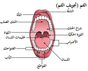
Illu mouth-ar.jpg 400 × 325; 44 KB
-
Illu mouth.jpg 400 × 325; 32 KB
-
Illu03 mouth zh.jpg 505 × 335; 79 KB
-
Illu03 mouth-ar.jpg 505 × 335; 88 KB
-
Illu03 mouth.jpg 505 × 335; 78 KB
-
Incisors Teeth (PSF).png 1.992 × 2.081; 768 KB
-
Insicors (PSF).png 2.494 × 1.769; 432 KB
-
InternationalJ.png 345 × 72; 3 KB
-
InternationalP.png 306 × 74; 2 KB
-
Jock Doe Chart.jpg 249 × 150; 19 KB
-
Kronenverlängerung.jpg 221 × 296; 22 KB
-
Kruemmungsmerkmal.png 700 × 250; 8 KB
-
Lower wisdom tooth neu.jpg 587 × 600; 25 KB
-
Mandibular 1st Molar.svg 520 × 370; 8 KB
-
MICAP-Befund.png 1.320 × 1.414; 541 KB
-
MIPAC-Chart.png 2.880 × 1.826; 733 KB
-
MIPAC.png 1.330 × 572; 64 KB
-
Molar Kaufläche.png 520 × 370; 76 KB
-
Molar relationship.jpg 472 × 371; 30 KB
-
OK UK zeichnung.gif 399 × 410; 22 KB
-
Optische Eigenschaften3 D 400.png 2.400 × 3.000; 190 KB
-
Outline drawing Mand1stMolar.jpg 1.992 × 1.479; 406 KB
-
Overjet-overbite.png 255 × 251; 16 KB
-
Palate 1 (PSF).png 1.703 × 1.674; 459 KB
-
PELL Y GREGORY.png 1.020 × 685; 548 KB
-
Periodontium.svg 339 × 665; 187 KB
-
Permanent maxillary Teeth - by Rokaya Yahia June 2015.jpg 2.776 × 2.640; 1,06 MB
-
Permanent-gebit.gif 676 × 513; 36 KB
-
Pierre Fauchard - Le Chirurgien Dentiste - dentures.png 504 × 902; 174 KB
-
Pitandfissurecaries01.PNG 427 × 293; 6 KB
-
Pitandfissurecaries02.PNG 427 × 293; 7 KB
-
Pitandfissurecaries03.PNG 427 × 293; 7 KB
-
Pitandfissurecaries04.PNG 427 × 293; 7 KB
-
Pitandfissurecaries05.PNG 427 × 293; 7 KB
-
Posseltb.png 566 × 582; 116 KB
-
Primary dentition.jpg 1.024 × 744; 114 KB
-
Prismemail.jpg 231 × 216; 8 KB
-
PrummelKlammer.jpg 216 × 310; 10 KB
-
Ptnadult.svg 420 × 210; 37 KB
-
Ptnchild.svg 420 × 210; 6 KB
-
Pulp cap.png 3.453 × 3.453; 2,44 MB
-
Pulp necrosis.jpg 1.600 × 1.600; 113 KB
-
Raspored ljudskih zuba.jpg 375 × 620; 36 KB
-
Runningroom.png 1.199 × 747; 55 KB
-
Schema dent dentine.png 512 × 798; 401 KB
-
Siebert 12 (jaws).jpg 1.302 × 1.667; 558 KB
-
Siebert 12.jpg 1.946 × 2.854; 1,33 MB
-
Sobo 1906 330.png 1.347 × 1.314; 5,07 MB
-
Sotokawazu01.jpg 505 × 435; 46 KB
-
Sotokawazu02.jpg 482 × 438; 39 KB
-
Sotokawazu29.jpg 503 × 488; 51 KB
-
Sotokawazu30.jpg 485 × 429; 52 KB
-
Sotokawazu31A.jpg 496 × 410; 47 KB
-
Sotokawazu32A.jpg 510 × 434; 46 KB
-
Sotokawazu33.jpg 433 × 493; 49 KB
-
Sotokawazu34.jpg 539 × 417; 59 KB
-
Sotokawazu35.jpg 495 × 401; 40 KB
-
Sotokawazu49.jpg 550 × 379; 55 KB
-
Sotokawazu50.jpg 550 × 417; 59 KB
-
Sotokawazu64.jpg 500 × 362; 42 KB
-
Sumter jane doe teeth chart.jpg 290 × 150; 22 KB
-
Tadasoto1.png 275 × 206; 15 KB
-
Tadasoto10.png 373 × 310; 19 KB
-
Tadasoto11.png 777 × 734; 58 KB
-
Tadasoto12.png 851 × 915; 73 KB
-
Tadasoto2.png 350 × 282; 20 KB
-
Tadasoto3.png 499 × 205; 17 KB
-
Tadasoto4.png 364 × 243; 20 KB
-
Tadasoto5.png 374 × 283; 18 KB
-
Tadasoto6.png 478 × 212; 16 KB
-
Tadasoto7.png 451 × 229; 18 KB
-
Tadasoto9.png 326 × 274; 18 KB
-
Teeth (PSF).png 1.605 × 1.566; 350 KB
-
Teeth model front FDI Notation.jpg 800 × 600; 98 KB
-
Teeth.JPG 596 × 800; 56 KB
-
TemporalTeethDeveploment.gif 351 × 504; 87 KB
-
The Dental cosmos (1890) (14775981321).jpg 2.208 × 1.440; 283 KB
-
The Dental cosmos (1890) (14776463311).jpg 1.636 × 2.324; 262 KB
-
The Dental cosmos (1912) (14766654424).jpg 2.464 × 1.396; 556 KB
-
The Periodontium.jpg 221 × 296; 14 KB
-
Tms-stift.PNG 335 × 428; 61 KB
-
Tooth 1 (PSF).png 1.077 × 1.872; 127 KB
-
Tooth 2 (PSF).png 1.850 × 1.508; 330 KB
-
Tooth Diagram.png 180 × 180; 21 KB
-
Tooth Picturewlabels.jpg 737 × 458; 92 KB
-
Toothbud annot.gif 359 × 302; 2,96 MB
-
Tuberculum carabelli.png 266 × 266; 48 KB
-
Tubérculo de Carabelli.jpg 266 × 266; 12 KB
-
Tönn.jpg 144 × 174; 6 KB
-
Winkelmerkmal.png 500 × 350; 7 KB
-
Wisdom Tooth (PSF).png 1.666 × 1.313; 103 KB
-
Wurzelmerkmal.png 500 × 350; 8 KB
-
Xray empty dental space new.jpg 285 × 240; 7 KB
-
Zahnaequator 78226 img.jpg 216 × 151; 6 KB
-
Zahneschema UK 1.jpg 799 × 600; 93 KB
-
Zahnfleischbluten.png 300 × 206; 12 KB
-
Zahnfleischentzuendung.png 300 × 206; 20 KB
-
Zahnformel Ferkel.png 231 × 100; 6 KB
-
Zahnformel Fohlen.png 231 × 100; 6 KB
-
Zahnformel Kaninchen.png 274 × 99; 6 KB
-
Zahnschema OK 1.jpg 800 × 600; 73 KB
-
Zahnseide co.png 365 × 373; 44 KB
-
Ανθρώπινο στόμα.jpg 800 × 780; 244 KB
-
Ротовая полость.jpg 302 × 250; 14 KB
-
Ротовая полость.png 400 × 325; 122 KB
-
Схема классификации СКЗ по топографии нахождения в зубной дуге согласно МКБ-10.jpg 3.866 × 2.766; 1,11 MB
-
ساختمان دندان.png 528 × 600; 122 KB


