[go: up one dir, main page]
More Web Proxy on the site http://driver.im/
You seem to have javascript disabled. Please note that many of the page functionalities won't work as expected without javascript enabled.


Evolution of Venom Systems

A topical collection in Toxins (ISSN 2072-6651). This collection belongs to the section "Animal Venoms".

Viewed by 260137

Editor


E-Mail Website
Collection Editor

Topical Collection Information

Dear Colleagues,

Venom systems are key evolutionary innovations used for competitor deterrence, defence and predation. The evolution of venom has been the trigger for the explosive radiation of many orders of animals. This Special Issue will be composed of papers exploring origin and diversification of venom systems and their intricate relationship with changes in predatory ecology. Reviews for this collection are on an invitation-only basis. Please contact Professor Fry to discuss ideas for a review so as to avoid overlap with other reviews.

Prof. Dr. Bryan Fry
Guest Editor

Manuscript Submission Information

Manuscripts for the topical collection can be submitted online at www.mdpi.com by registering and logging in to this website. Once you are registered, click here to go to the submission form. All papers will be peer-reviewed. Accepted papers will be published continuously in the journal (as soon as accepted) and will be listed together on this website. The topical collection considers regular research articles, short communications and review articles. A guide for authors and other relevant information for submission of manuscripts is available on the Instructions for Authors page.

Please visit the Instructions for Authors page before submitting a manuscript. The article processing charge (APC) for publication in this open access journal is 2700 CHF (Swiss francs).

Published Papers (22 papers)

2020

Jump to: 2018, 2017, 2016, 2015, 2014, 2013

25 pages, 3265 KiB  
Article
Size Matters: An Evaluation of the Molecular Basis of Ontogenetic Modifications in the Composition of Bothrops jararacussu Snake Venom
by Luciana A. Freitas-de-Sousa, Pedro G. Nachtigall, José A. Portes-Junior, Matthew L. Holding, Gunnar S. Nystrom, Schyler A. Ellsworth, Noranathan C. Guimarães, Emilly Tioyama, Flora Ortiz, Bruno R. Silva, Tobias S. Kunz, Inácio L. M. Junqueira-de-Azevedo, Felipe G. Grazziotin, Darin R. Rokyta and Ana M. Moura-da-Silva
Toxins 2020, 12(12), 791; https://doi.org/10.3390/toxins12120791 - 11 Dec 2020
Cited by 24 | Viewed by 4963
Abstract
Ontogenetic changes in venom composition have been described in Bothrops snakes, but only a few studies have attempted to identify the targeted paralogues or the molecular mechanisms involved in modifications of gene expression during ontogeny. In this study, we decoded B. jararacussu venom [...] Read more.
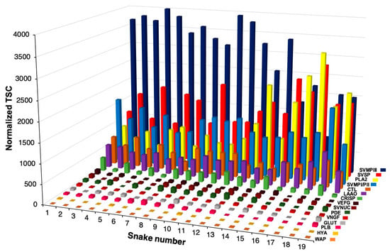
Ontogenetic changes in venom composition have been described in Bothrops snakes, but only a few studies have attempted to identify the targeted paralogues or the molecular mechanisms involved in modifications of gene expression during ontogeny. In this study, we decoded B. jararacussu venom gland transcripts from six specimens of varying sizes and analyzed the variability in the composition of independent venom proteomes from 19 individuals. We identified 125 distinct putative toxin transcripts, and of these, 73 were detected in venom proteomes and only 10 were involved in the ontogenetic changes. Ontogenetic variability was linearly related to snake size and did not correspond to the maturation of the reproductive stage. Changes in the transcriptome were highly predictive of changes in the venom proteome. The basic myotoxic phospholipases A2 (PLA2s) were the most abundant components in larger snakes, while in venoms from smaller snakes, PIII-class SVMPs were the major components. The snake venom metalloproteinases (SVMPs) identified corresponded to novel sequences and conferred higher pro-coagulant and hemorrhagic functions to the venom of small snakes. The mechanisms modulating venom variability are predominantly related to transcriptional events and may consist of an advantage of higher hematotoxicity and more efficient predatory function in the venom from small snakes. Full article
Show Figures

Figure 1

Figure 1
<p>Distribution of the toxin families in the venom of 19 specimens of <span class="html-italic">Bothrops jararacussu</span> identified from proteome analysis. Relative expression is indicated by the normalized Total Spectrum Count (TSC) in each snake. CTL (C-Type Lectin), PLA<sub>2</sub> (Phospholipase A<sub>2</sub>), SVMP classes PI, PII, and PIII (Snake Venom Metalloproteinase), SVSP (Snake Venom Serine Protease), VEGF (Vascular Endothelial Growth Factor), LAAO (L-amino Acid Oxidase), CRISP (Cysteine-rich secretory protein), HYAL (Hyaluronidase), VNGF (Venom Nerve Growth Factor), SVNUC (Snake Venom Nucleotidase), PDE (Venom Phosphodiesterase), PLB (Phospholipase B) and GLUT (Snake Venom Glutaminyl Cyclase). The higher the snake number, the greater the size of the snake.</p>
Full article ">Figure 2
<p>Correlation of the main toxin families of <span class="html-italic">Bothrops jararacussu</span> venom with: (<b>A</b>) the individuals’ snout-vent length (SVL), (<b>B</b>) reproductive status, sex, and place of collection. Relative expression is indicated by the normalized Total Spectrum Count (TSC) in each snake. CTL (C-Type Lectin), PLA<sub>2</sub> (Phospholipase A<sub>2</sub>), SVMP classes PI, PII and PIII (Snake Venom Metalloproteinase), SVSP (Snake Venom Serine Protease), LAAO (L-amino Acid Oxidase), CRISP (Cysteine-rich secretory protein). * <span class="html-italic">p</span> &lt; 0.05.</p>
Full article ">Figure 3
<p>Distribution of PLA<sub>2</sub> isoforms present in the venoms of 19 individuals from <span class="html-italic">B. jararacussu</span> and functional inferences. Mature deduced protein sequences of PLA<sub>2</sub> were aligned together with sequences of PLA<sub>2</sub>s isolated from other venoms with functions well-established by previous studies using experimental approaches, as follows: a non-toxic phospholipase A2 from the venom of <span class="html-italic">Notechis scutatus scutatus</span>; Bothropstoxin-1 (Q90249.3), Bothropstoxin-2 (P45881) and BthAI (Q8AXY1.1), <span class="html-italic">B. jararacussu</span> venom; acidic PLA<sub>2</sub> (Q2HZ28.1), <span class="html-italic">B. erythromelas</span> venom. <span class="html-italic">Notechis</span> PLA<sub>2</sub> (P08873.1) sequence was used to root the tree. The maximum likelihood phylogenetic tree was generated using RaxML (v8.2.12; Stamatakis 2014). The heatmap on the right indicates the number of normalized Exclusive Unique Spectrum Counts (EUSC) of MS/MS in each venom sample, which are as indicated at the top of the figure. Bootstrap values are described only when greater than 75.</p>
Full article ">Figure 4
<p>Distribution of SVMPIII isoforms present in the venoms of 19 individuals from <span class="html-italic">B. jararacussu</span> and functional inferences. Mature deduced protein sequences of SVMPIII were aligned together with sequences of SVMPs isolated from other venoms with functions well-established by previous studies using experimental approaches, as follows: BjussuMP-1P (Q1PHZ4; from <span class="html-italic">B. jararacussu</span> venom; Batroxrhagin, <span class="html-italic">Bothrops atrox</span> venom (ALB00542.1); hemorrhagic factor 3 (HF3), <span class="html-italic">B. jararaca</span> venom (Q98UF9.3); vascular apoptosis-inducing protein 1 (VAP-1), <span class="html-italic">Crotalus atrox</span> venom (Q9DGB9.1); berythractivase, <span class="html-italic">B. erythromelas</span> venom (Q8UVG0.1); VaF1, <span class="html-italic">Vipera ammodytes ammodytes</span> venom (AJC52543). The disintegrin and metalloproteinase domains of <span class="html-italic">Mus musculus</span> ADAM28 (NP034212.1) was used to root the tree. The maximum likelihood phylogenetic tree was generated using RaxML (v8.2.12; Stamatakis 2014). The heatmap on the right indicates the number of normalized Exclusive Unique Spectrum Counts (EUSC) of MS/MS in each venom sample, which are as indicated at the top of the figure. Bootstrap values are described only when greater than 75.</p>
Full article ">Figure 5
<p>Comparisons of gene transcription (TMM normalized) rates in the venom gland and protein abundance (normalized EUSC—Exclusive Unique Spectrum Count) in the venom of individual <span class="html-italic">B. jararacussu</span> snakes. Each spot represents detected transcription and translation levels for a specific toxin family from one specimen. Dotted lines indicate a hypothetical correspondence between transcript and protein abundances.</p>
Full article ">Figure 6
<p>Comparison between levels of transcripts (TMM normalized) and protein abundance (normalized EUSC—Exclusive Unique Spectrum Count) of the isoforms PLA002, SVMPIII002, and SVMPIII009 in the venoms of <span class="html-italic">B. jararacussu</span> snakes. Dotted lines indicate a hypothetical correspondence between transcript and protein abundances.</p>
Full article ">Figure 7
<p>Functional activities of <span class="html-italic">Bothrops jararacussu</span> venom samples. (<b>A</b>)—Metalloproteinase activity (SVMP) was measured by fluorometric assays using the substrate Abz-AGLA-EDDnp. (<b>B</b>)—Phospholipase A<sub>2</sub> (PLA<sub>2</sub>) activity was tested using the 4-nitro-3- [octanoyloxy] benzoic acid substrate. (<b>C</b>)—Procoagulant activity was evaluated by the amount of venom (µg) capable of inducing the coagulation of human citrated plasma in up to 60 s and is represented by the reciprocal Minimum Coagulant Dose (1/MCD). (<b>D</b>)—For hemorrhagic activity, 10 µg of each pool of venoms were injected in the dorsum of mice, and the hemorrhagic lesions were measured three hours after injection. (<b>E</b>)—The myotoxic activity was assayed using 50 µg of pools of venoms injected intramuscularly into the gastrocnemius muscle in Swiss mice, and after 3 h, the sera were assayed for creatine–kinase activity. (<b>F</b>)—For the evaluation of lethal activity, samples containing 200 μg of each venom in a final volume of 200 μL were injected intraperitoneally into groups of four mice. Phosphate-buffered saline (PBS)—was the control in all in vivo experiments. The results are representative of two independent experiments. <b>*</b> <span class="html-italic">p</span> &lt; 0.05.</p>
Full article ">
2018

Jump to: 2020, 2017, 2016, 2015, 2014, 2013

2 pages, 216 KiB  
Addendum
Addendum: Aird, S.D. et al. Coralsnake Venomics: Analyses of Venom Gland Transcriptomes and Proteomes of Six Brazilian Taxa. Toxins 2017, 9(6), 187
by Steven D. Aird, Nelson Jorge Da Silva, Lijun Qiu, Alejandro Villar-Briones, Vera Aparecida Saddi, Mariana Pires de Campos Telles, Miguel L. Grau and Alexander S. Mikheyev
Toxins 2018, 10(5), 172; https://doi.org/10.3390/toxins10050172 - 24 Apr 2018
Viewed by 3744
Abstract
Following publication of this paper, Dr. Daniel Dashevsky discovered to our chagrin, that the transcriptomic datasets uploaded to the DNA Databank of Japan (DDBJ) contained numerous complete 3FTx sequences that were not included in our paper.[...] Full article

2017

Jump to: 2020, 2018, 2016, 2015, 2014, 2013

14820 KiB  
Article
Enter the Dragon: The Dynamic and Multifunctional Evolution of Anguimorpha Lizard Venoms
by Ivan Koludarov, Timothy NW Jackson, Bianca op den Brouw, James Dobson, Daniel Dashevsky, Kevin Arbuckle, Christofer J. Clemente, Edward J. Stockdale, Chip Cochran, Jordan Debono, Carson Stephens, Nadya Panagides, Bin Li, Mary-Louise Roy Manchadi, Aude Violette, Rudy Fourmy, Iwan Hendrikx, Amanda Nouwens, Judith Clements, Paolo Martelli, Hang Fai Kwok and Bryan G. Fryadd Show full author list remove Hide full author list
Toxins 2017, 9(8), 242; https://doi.org/10.3390/toxins9080242 - 6 Aug 2017
Cited by 38 | Viewed by 31895
Abstract
While snake venoms have been the subject of intense study, comparatively little work has been done on lizard venoms. In this study, we have examined the structural and functional diversification of anguimorph lizard venoms and associated toxins, and related these results to dentition [...] Read more.
While snake venoms have been the subject of intense study, comparatively little work has been done on lizard venoms. In this study, we have examined the structural and functional diversification of anguimorph lizard venoms and associated toxins, and related these results to dentition and predatory ecology. Venom composition was shown to be highly variable across the 20 species of Heloderma, Lanthanotus, and Varanus included in our study. While kallikrein enzymes were ubiquitous, they were also a particularly multifunctional toxin type, with differential activities on enzyme substrates and also ability to degrade alpha or beta chains of fibrinogen that reflects structural variability. Examination of other toxin types also revealed similar variability in their presence and activity levels. The high level of venom chemistry variation in varanid lizards compared to that of helodermatid lizards suggests that venom may be subject to different selection pressures in these two families. These results not only contribute to our understanding of venom evolution but also reveal anguimorph lizard venoms to be rich sources of novel bioactive molecules with potential as drug design and development lead compounds. Full article
Show Figures

Figure 1

Figure 1
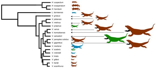
<p>Organismal relationships, sizes and ecological niches occupied (blue = aquatic, brown = terrestrial, green = arboreal). Phylogeny based on [<a href="#B65-toxins-09-00242" class="html-bibr">65</a>,<a href="#B66-toxins-09-00242" class="html-bibr">66</a>,<a href="#B67-toxins-09-00242" class="html-bibr">67</a>]. Note that as it only includes species from this study, it excludes the other anguimorph lizards such as anguids that intervene between <span class="html-italic">Heloderma</span> and <span class="html-italic">Lanthantus/Varanus.</span> Red lineages are terrestrial, green are arboreal, and blue are aquatic.</p>
Full article ">Figure 2
<p>Scanning electron microscopy showing the (<b>A</b>) plesiomorphic moderate/medium anterior and posterior serrations such as in <span class="html-italic">V. giganteus</span>, (<b>B</b>) the derived non-serrated condition as in <span class="html-italic">V. mertensi</span>, (<b>B</b>) or (<b>C</b>,<b>D</b>) derived deep anterior and posterior serrations as in <span class="html-italic">V. komodoensis</span>.</p>
Full article ">Figure 3
<p>Ancestral state reconstructions over branches for anterior teeth serrations and posterior teeth serrations inhibition where warmer colours represent more serrations. Bars indicate 95% confidence intervals for the estimate at each node. Note that due to the high dynamicity of venom evolution these quickly become broad as you move down the tree. Phylogeny follows [<a href="#B65-toxins-09-00242" class="html-bibr">65</a>,<a href="#B66-toxins-09-00242" class="html-bibr">66</a>,<a href="#B67-toxins-09-00242" class="html-bibr">67</a>].</p>
Full article ">Figure 4
<p>1D gel comparisons: (<b>A</b>) Vmer = <span class="html-italic">V. mertensi</span>, Vgig = <span class="html-italic">V. giganteus</span>, Vrub = <span class="html-italic">V. panoptes rubidus</span>, Vmit = <span class="html-italic">V. mitchelli</span>, Vsca = <span class="html-italic">V. scalaris</span>, Vtri = <span class="html-italic">V. tristis</span>; (<b>B</b>) Vgil = <span class="html-italic">V. gilleni</span>, Vaca = <span class="html-italic">V. acanthurus</span>, Vbar = <span class="html-italic">V. baritji</span>; (<b>C</b>) Vgri = <span class="html-italic">V. griseus</span>, Vpra = <span class="html-italic">V. prasinus</span>, Vjob = <span class="html-italic">V. jobiensis</span>, Vmel = <span class="html-italic">V. melinus</span>, Vsal = <span class="html-italic">V. salvadorii</span>, Vvar = <span class="html-italic">V. varius</span>; and (<b>D</b>) Hsus = <span class="html-italic">H. suspectum</span>, Lbor = <span class="html-italic">L. borneensis.</span> Molecular markers are shown in kDa.</p>
Full article ">Figure 5
<p>Varanid venom 2D gel comparisons showing the relative kallikrein presence. (<b>A</b>) <span class="html-italic">Varanus acanthurus</span>, (<b>B</b>) <span class="html-italic">Varanus giganteus</span>, (<b>C</b>) <span class="html-italic">Varanus gilleni</span>, (<b>D</b>) <span class="html-italic">Varanus griseus</span>, (<b>E</b>) <span class="html-italic">Varanus mertensi</span>, (<b>F</b>) <span class="html-italic">Varanus mitchelli</span>, (<b>G</b>) <span class="html-italic">Varanus prasinus</span>, (<b>H</b>) <span class="html-italic">Varanus rubidus</span>, (<b>I</b>) <span class="html-italic">Varanus salvadorii</span>, (<b>J</b>) <span class="html-italic">Varanus scalaris</span>, (<b>K</b>) <span class="html-italic">Varanus tristis</span>, and (<b>L</b>) <span class="html-italic">Varanus varius</span>.</p>
Full article ">Figure 6
<p>Molecular phylogenetic relationships of anguimorph lizard kallikrein enzymes. Numbers at node indicate probability. A monophyletic clade of toxicoferan mandibular and maxillary gland sequences is shown in green.</p>
Full article ">Figure 7
<p>Three dimensional structure of a kallikrein toxin coloured according to (<b>A</b>) AL2CO amino acid conservation score (conserved sites in teal and variable sites in orange), (<b>B</b>) FUBAR strength of persistent selection (sites under purifying selection in blue and sites under diversifying selection in red), and (<b>C</b>) MEME significance levels for episodes of diversifying selection during the evolution of the toxin family (moderately significant sites in dark green, highly significant sites in light green, and extremely significant sites in yellow).</p>
Full article ">Figure 8
<p>Molecular phylogenetic relationships of anguimorph lizard lysosomal acid lipase enzymes relative to other sequences. Varanid venom gland sequences are shown in green.</p>
Full article ">Figure 9
<p>Activity levels for the bioactivity assays shown as <span class="html-italic">n</span> = 3 means with standard deviation error bars.</p>
Full article ">Figure 10
<p>Ancestral state reconstruction of serine protease substrate activity, where warmer colours represent more potent substrate cleavage. Bars indicate 95% confidence intervals for the estimate at each node. Note that due to the high dynamicity of venom evolution the ranges quickly become broad as one moves down the tree. Phylogeny follows [<a href="#B65-toxins-09-00242" class="html-bibr">65</a>,<a href="#B66-toxins-09-00242" class="html-bibr">66</a>,<a href="#B67-toxins-09-00242" class="html-bibr">67</a>].</p>
Full article ">Figure 11
<p>Fibrinogen cleavage over time. For <b>gels</b> (<b>A</b>–<b>H</b>) lanes 1&amp;6 = time zero, lanes 2&amp;7 = 1 min incubation, lanes 3&amp;8 = 5 min incubation, lanes 4&amp;9 = 20 min incubation, and lanes 5&amp;10 = 60 min incubation. For <b>gel</b> (<b>I</b>) lanes 1&amp;6 = time zero, lanes 2, 4, 7 &amp; 9 = 5 min incubation, and lanes 3, 5, 8, &amp; 10 = 60 min incubation. L. born = <span class="html-italic">L. borneensis</span>, H. exas = <span class="html-italic">H. exasperatum</span>, H. horr = <span class="html-italic">H. horridum</span>, H. susp = <span class="html-italic">H. suspectum</span>, V. acan = <span class="html-italic">V. acanthurus</span>, V. bari = <span class="html-italic">V. baritji</span>, V. giga = <span class="html-italic">V. giganteus</span>, Vgl = <span class="html-italic">V. gilleni</span>, V. gris = <span class="html-italic">V. griseus</span>, V. komo = <span class="html-italic">V. komodoensis</span>, V. jobi = <span class="html-italic">V. jobiensis</span>, V. miti = <span class="html-italic">V. mitchelli</span>, VmL = <span class="html-italic">V. melinus</span>, Vme = <span class="html-italic">V. mertensi</span>, V. pras = <span class="html-italic">V. prasinus</span>, Vru = <span class="html-italic">V. panoptes rubidus</span>, V. scal = <span class="html-italic">V. scalaris</span>, V. salv = <span class="html-italic">V. salvadorii</span>, V. tris = <span class="html-italic">V. tristis</span>, and V. vari = <span class="html-italic">V. varius</span>. Gels were run in triplicate, with the patterns congruent between replicates.</p>
Full article ">Figure 12
<p>Relative cleavage of alpha (red) or beta (blue) chains of fibrinogen. X-axis is time (min) and y-axis is percentage of intact chain remaining. Error bars indicate <span class="html-italic">n</span> = 3 standard deviation.</p>
Full article ">Figure 13
<p>Ancestral state reconstructions over branches for destructive cleavage of fibrinogen alpha and beta chains. Bars indicate 95% confidence intervals for the estimate at each node. Note that due to the high dynamicity of venom evolution these quickly become broad as one moves down the tree. Phylogeny follows [<a href="#B65-toxins-09-00242" class="html-bibr">65</a>,<a href="#B66-toxins-09-00242" class="html-bibr">66</a>,<a href="#B67-toxins-09-00242" class="html-bibr">67</a>].</p>
Full article ">Figure 14
<p>Ancestral state reconstructions over branches comparing substrate consumption relative to alpha chain destruction. Bars indicate 95% confidence intervals for the estimate at each node. Note that due to the high dynamicity of venom evolution these quickly become broad as one moves down the tree. Phylogeny follows [<a href="#B65-toxins-09-00242" class="html-bibr">65</a>,<a href="#B66-toxins-09-00242" class="html-bibr">66</a>,<a href="#B67-toxins-09-00242" class="html-bibr">67</a>].</p>
Full article ">Figure 15
<p>Ancestral state reconstructions over branches comparing substrate consumption relative to beta chain destruction. Bars indicate 95% confidence intervals for the estimate at each node. Note that due to the high dynamicity of venom evolution these quickly become broad as one moves down the tree. Phylogeny follows [<a href="#B65-toxins-09-00242" class="html-bibr">65</a>,<a href="#B66-toxins-09-00242" class="html-bibr">66</a>,<a href="#B67-toxins-09-00242" class="html-bibr">67</a>].</p>
Full article ">Figure 16
<p>Ancestral state reconstructions over branches for PLA<sub>2</sub> enzyme substrate consumption where warmer colours represent more fluorescence production. Bars indicate 95% confidence intervals for the estimate at each node. Note that due to the high dynamicity of venom evolution these quickly become broad as one moves down the tree. Phylogeny follows [<a href="#B65-toxins-09-00242" class="html-bibr">65</a>,<a href="#B66-toxins-09-00242" class="html-bibr">66</a>,<a href="#B67-toxins-09-00242" class="html-bibr">67</a>].</p>
Full article ">Figure 17
<p>Change in rat ileum smooth muscle contractility after administration of crude 50 μg/mL <span class="html-italic">V. varius</span> venom. On the Y-axis is the pulling force in relative units; on the X-axis is time. Experiment was run in triplicate and results were congruent between replicates.</p>
Full article ">
19648 KiB  
Article
Coralsnake Venomics: Analyses of Venom Gland Transcriptomes and Proteomes of Six Brazilian Taxa
by Steven D. Aird, Nelson Jorge Da Silva, Lijun Qiu, Alejandro Villar-Briones, Vera Aparecida Saddi, Mariana Pires de Campos Telles, Miguel L. Grau and Alexander S. Mikheyev
Toxins 2017, 9(6), 187; https://doi.org/10.3390/toxins9060187 - 8 Jun 2017
Cited by 69 | Viewed by 9880
Abstract
Venom gland transcriptomes and proteomes of six Micrurus taxa (M. corallinus, M. lemniscatus carvalhoi, M. lemniscatus lemniscatus, M. paraensis, M. spixii spixii, and M. surinamensis) were investigated, providing the most comprehensive, quantitative data on Micrurus venom [...] Read more.
Venom gland transcriptomes and proteomes of six Micrurus taxa (M. corallinus, M. lemniscatus carvalhoi, M. lemniscatus lemniscatus, M. paraensis, M. spixii spixii, and M. surinamensis) were investigated, providing the most comprehensive, quantitative data on Micrurus venom composition to date, and more than tripling the number of Micrurus venom protein sequences previously available. The six venomes differ dramatically. All are dominated by 2–6 toxin classes that account for 91–99% of the toxin transcripts. The M. s. spixii venome is compositionally the simplest. In it, three-finger toxins (3FTxs) and phospholipases A2 (PLA2s) comprise >99% of the toxin transcripts, which include only four additional toxin families at levels ≥0.1%. Micrurus l. lemniscatus venom is the most complex, with at least 17 toxin families. However, in each venome, multiple structural subclasses of 3FTXs and PLA2s are present. These almost certainly differ in pharmacology as well. All venoms also contain phospholipase B and vascular endothelial growth factors. Minor components (0.1–2.0%) are found in all venoms except that of M. s. spixii. Other toxin families are present in all six venoms at trace levels (<0.005%). Minor and trace venom components differ in each venom. Numerous novel toxin chemistries include 3FTxs with previously unknown 8- and 10-cysteine arrangements, resulting in new 3D structures and target specificities. 9-cysteine toxins raise the possibility of covalent, homodimeric 3FTxs or heterodimeric toxins with unknown pharmacologies. Probable muscarinic sequences may be reptile-specific homologs that promote hypotension via vascular mAChRs. The first complete sequences are presented for 3FTxs putatively responsible for liberating glutamate from rat brain synaptosomes. Micrurus C-type lectin-like proteins may have 6–9 cysteine residues and may be monomers, or homo- or heterodimers of unknown pharmacology. Novel KSPIs, 3× longer than any seen previously, appear to have arisen in three species by gene duplication and fusion. Four species have transcripts homologous to the nociceptive toxin, (MitTx) α-subunit, but all six species had homologs to the β-subunit. The first non-neurotoxic, non-catalytic elapid phospholipase A2s are reported. All are probably myonecrotic. Phylogenetic analysis indicates that the six taxa diverged 15–35 million years ago and that they split from their last common ancestor with Old World elapines nearly 55 million years ago. Given their early diversification, many cryptic micrurine taxa are anticipated. Full article
Show Figures

Graphical abstract

Graphical abstract
Full article ">Figure 1
<p>These Brazilian <span class="html-italic">Micrurus</span> venoms all contain three-finger toxins (3FTxs) and phospholipases A<sub>2</sub> (PLA<sub>2</sub>s), but but they vary greatly in the relative proportions and subclasses thereof, and in the types and amounts of minor toxins as well. (<b>A</b>) Major toxins comprising ≥2% of the toxin portion a given transcriptome. The “other” portion of each venom (black) was comprised of minor components; (<b>B</b>) Minor toxins representing between 0.1% and 2.0% of the toxin transcriptome. Each venom contained still other toxins at trace levels, each amounting to less than 0.1% of the transcriptome. <span class="html-italic">Micrurus s. spixii</span> possesses the simplest venom, with 3FTxs and PLA<sub>2</sub>s accounting for just over 99% of the transcriptome, and comprising only six major and minor toxin classes.</p>
Full article ">Figure 2
<p><span class="html-italic">Micrurus</span> venoms are rich in 3FTxs displaying an astonishing variety of primary structures. Micrurine 3FTxs may possess 8, 9, or 10 Cys residues with different disulfide patterns in each group. Pharmacologies are almost entirely unknown. Some probably target nicotinic acetylcholine receptors of reptilian neuromuscular junctions, but their potential targets in mammals are unknown. Most 3FTxs have 21-residue signal peptides. In the interest of creating a figure of manageable size for the journal format, all signal peptides have been deleted here, and only sequences originating in this study have been included. Full sequences of all 184 micrurine 3FTxs can be found in <a href="#app1-toxins-09-00187" class="html-app">Figure S2</a>.</p>
Full article ">Figure 3
<p>Primary and 3D structures of 9-Cys 3FTxs from <span class="html-italic">Micrurus</span> venoms include two structural subclasses. (<b>A</b>) To date, micrurine 3FTxs with 9 cysteines have been found only in venoms of <span class="html-italic">Micrurus l. lemniscatus</span> and <span class="html-italic">M. altirostris</span>; however, among these, the extra cysteine can appear in either of two positions, indicated by red arrows. Putative conserved disulfide bonds are indicated with black bars. Signal peptides, 21 residues in length, were almost invariant (MKTLL LTLVV VTIVC LDFGH T). <span class="html-italic">M. l. lemniscatus</span> toxin DN120340 had an L/Q substitution in position 4 and a V/L substitution in position 14; (<b>B</b>) Front and side views of the ribbon model of <span class="html-italic">M. l. lemniscatus</span> DN120340; (<b>C</b>) Front and side views of the ribbon model of <span class="html-italic">M. l. lemniscatus</span> DN120555. SWISS-MODEL was used to select the best templates for the <span class="html-italic">Micrurus</span> toxins (<span class="html-italic">Laticauda semifasciata</span>, erabutoxin, 2era.1.A for <span class="html-italic">M. l. lemnsicatus</span> DN120340 and <span class="html-italic">Naja atra</span> cobrotoxin (1coe.1.A) for <span class="html-italic">M. l. lemniscatus</span> DN120555) and to construct a preliminary model. Then models were refined and energy minimizations were performed with UCSF Chimera [<a href="#B21-toxins-09-00187" class="html-bibr">21</a>]. Disulfide bonds are shown in white.</p>
Full article ">Figure 4
<p>Micrurine 3FTxs with 10 cysteine residues include several subclasses. (<b>A</b>) 3FTxs with 10 cysteines show the disulfide bond pattern of γ-bungarotoxin (red bar), but not of α- and κ-bungarotoxins (black bar). Thus, homologs of the latter two 3FTx subclasses probably are not found in New World coralsnake venoms. Residues to the left of the black vertical line constitute signal peptides; (<b>B</b>) Ribbon models of three different subclasses of 10-Cys 3FTxs from the venom of <span class="html-italic">M. paraensis</span>: DN85120 (tan), DN85432 (blue), and DN86421 (violet), from (<b>A</b>). 3D representations were made with UCSF Chimera [<a href="#B21-toxins-09-00187" class="html-bibr">21</a>] based on models generated with SWISS-MODEL, using the structure of candoxin (1jgk.1.A) as a template for DN85120, bucandin (1ijc.1.A) as a template for DN85432, both from <span class="html-italic">Bungarus candidus</span> venom, and <span class="html-italic">Bungarus multicinctus</span> γ-bungarotoxin (1mr6.1.A) for DN86421. Disulfide bonds are shown in white.</p>
Full article ">Figure 5
<p>3FTxs bearing 8 cysteine residues show two very different disulfide bond patterns. (<b>A</b>) The classic short α-neurotoxin/cardiotoxin pattern, represented above by <span class="html-italic">Naja nigricollis</span> Cardiotoxin 1 and by <span class="html-italic">Micrurus fulvius</span> 3FTxs 9a &amp; b, is employed by all but two micrurine 3FTxs (<span class="html-italic">M. tener</span> 3FTx1 and <span class="html-italic">M. fulvius</span> 3FTx5a). The novel disulfide in the latter two toxins is indicated with a red bar. Conserved disulfides are denoted with black bars. Signal peptides are shown to the left of the black vertical line; (<b>B</b>) Ribbon models of the two subclasses of 8-Cys 3FTxs from the venom of <span class="html-italic">M. fulvius</span>: 3FTx 5a (JAB52850.1) (tan) and 3FTx 9a (JAS05068.1) (blue) from (<b>A</b>). Disulfide bonds are shown in white. SWISS-MODEL selected bucandin (1f94.1.A) from <span class="html-italic">Bungarus candidus</span> venom as the best template for <span class="html-italic">M. fulvius</span> 3FTx 5a, and 3FTx 3b (4rud.1.B) from <span class="html-italic">M. fulvius</span> venom as the best template for <span class="html-italic">M. fulvius</span> 3FTx 9a. Models were refined and energy minimizations were performed using UCSF Chimera [<a href="#B21-toxins-09-00187" class="html-bibr">21</a>].</p>
Full article ">Figure 6
<p>To date, all <span class="html-italic">Micrurus</span> venoms contain 3FTxs with significant similarities to muscarinic toxins from <span class="html-italic">Dendroaspis angusticeps</span> venom; however, these toxins also display numerous differences. It seems unlikely that these toxins could exhibit the same pharmacology in mammals as the mamba toxins, but they may antagonize mAChRs in reptiles, fishes, and onychophorans, the natural prey of coralsnakes. Aligned sequences of mamba muscarinic antagonists and putative muscarinic toxins from <span class="html-italic">Micrurus</span> venoms. Signal peptides are shown to the left of the black vertical line.</p>
Full article ">Figure 7
<p>Freire Donato [<a href="#B68-toxins-09-00187" class="html-bibr">68</a>] determined the N-terminal amino acid sequence of a toxin from the venom of <span class="html-italic">M. l. carvalhoi</span> that provokes the release of glutamate from rat cortical synaptosomes. While that sequence contained two cysteine residues that appear spurious, when those are deleted, her sequence aligns well with sequences from five of the six <span class="html-italic">Micrurus</span> transcriptomes. These appear to represent the first complete sequences of snake toxins that provoke glutamate release.</p>
Full article ">Figure 8
<p>The galactose-binding lectin from <span class="html-italic">Micrurus corallinus</span> venom (DN79899_c0_g1_i1|m30755) showing the molecule’s secondary structure (left) and the same model with the aromatic residues shown in a space-filling representation. Aromatic residues tend to be clustered in the core, but most tyrosines have their hydroxyl groups exposed on the surface and several of the tryptophans are also partially exposed. The functional reason for this extremely high aromatic content is unknown. The model was made with SWISS-MODEL using a GBL from <span class="html-italic">Protobothrops mucrosquamatus</span> venom as a template. The model was then further refined and energy minimization was performed using Chimera [<a href="#B21-toxins-09-00187" class="html-bibr">21</a>].</p>
Full article ">Figure 9
<p>Four of the species examined here have sequences that are strikingly similar to the α subunit of the nociceptive toxin (MitTx) from <span class="html-italic">Micrurus tener</span> venom, which even the closely related <span class="html-italic">M. fulvius</span> lacks. Nonetheless, the South American <span class="html-italic">Micrurus</span> toxins have five substitutions (indicated by triangles), four of which are strongly non-synonymous. Signal peptides 24 residues long were essentially invariant (MSSGG LLLLL GLLTL CAELT PVSS). Only <span class="html-italic">M. l. lemniscatus</span> toxin DN110178 had a G/R substitution in position 5.</p>
Full article ">Figure 10
<p>All six <span class="html-italic">Micrurus</span> species examined produced a close homolog of MitTxβ, the nociceptive toxin β subunit from <span class="html-italic">Micrurus tener</span>, including <span class="html-italic">M. paraensis</span> and <span class="html-italic">M. l. carvalhoi</span>, which apparently do not produce the α-subunit. The South American <span class="html-italic">Micrurus</span> sequences are basically identical, and differ from that of MitTxβ at only six positions (indicated by triangles). The three M. tener PLA<sub>2</sub> sequences had 21-residue signal peptides while MitTxβ and putative homologs had 24-residue signal peptides. 21-residue signal peptides had the sequence MNPAH LLVLA AVCVS LLGASS. MitTxβ and putative homologs had a three-residue N-terminal extension, MDK.</p>
Full article ">Figure 11
<p>When <span class="html-italic">Micrurus</span> species for which transcriptomic data are available, are arranged in an approximately northwestern to southeastern sequence, the gradient of high PLA<sub>2</sub> and low 3FTx concentrations in the North to high 3FTx and low PLA<sub>2</sub> concentration in the South previously suggested by Fernández et al. [<a href="#B170-toxins-09-00187" class="html-bibr">170</a>], no longer appears as obvious.</p>
Full article ">Figure 12
<p>Structures of 121 new micrurine phospholipases A<sub>2</sub>, illustrating their great structural diversity. For a comparison of all known micrurine PLA2s, see <a href="#app1-toxins-09-00187" class="html-app">Figure S22</a>. (<b>A</b>) Of 121 PLA<sub>2</sub>s with partial or complete structures, 202 are apparently catalytic, having the requisite H48, D49, Y52, and D101 in their active sites. The remaining 42 are apparently non-catalytic. Positions indicated in red across the bottom designate residues involved in Ca<sup>2+</sup>-binding [<a href="#B172-toxins-09-00187" class="html-bibr">172</a>]. Positions indicated in blue are catalytic residues. The anticoagulant site [<a href="#B173-toxins-09-00187" class="html-bibr">173</a>] and the neurotoxic hydrophobic region [<a href="#B172-toxins-09-00187" class="html-bibr">172</a>,<a href="#B174-toxins-09-00187" class="html-bibr">174</a>] are indicated by purple and green bars, respectively. Residues proposed by Alape-Girón et al. [<a href="#B175-toxins-09-00187" class="html-bibr">175</a>] to be involved in myotoxicity of elapid PLA<sub>2</sub>s are indicated below with black cells. Residues participating in the hydrophobic channel, via which substrate enters the catalytic site are indicated in gray [<a href="#B176-toxins-09-00187" class="html-bibr">176</a>]. PLA<sub>2</sub>s from the two North American species, <span class="html-italic">M. fulvius</span> and <span class="html-italic">M. tener</span>, form a structural cluster that is relatively distinctive from the much more variable South American sequences, which probably also represent much greater pharmacological diversity. Numbers of cysteine residues are given for sequences that are complete. Numbers of cysteines in parentheses are for sequences truncated at the N-terminal end, for which the cysteines can be reliably predicted, owing to the relative invariability of the N-termini. Asterisks indicate stop codons.Micrurine venoms include 42 presumably non-catalytic PLA<sub>2</sub>s that appear to be myotoxic. These comprise four loose subclasses, shown here separated by horizontal lines. The first two groups possess all four catalytic residues, but apparently have a disrupted Ca<sup>2+</sup>-binding site (D or I in lieu of G30, and R in place of G32). Toxins in the third group have H48, but D49 has been replaced by Y49 or F49, and G30 with N or S. The fourth subclass, comprising 9 PLA<sub>2</sub>s from all six Brazilian species and <span class="html-italic">M. tener</span>, has replaced H48 with Q and D49 with K. G30 has been replaced with D, I, or V. Thus, it seems improbable that any of these can bind Ca<sup>2+</sup>; (<b>B</b>) Space-filling models of catalytic residues, Ca<sup>2+</sup>-binding residues, and residues forming the hydrophobic channels of four micrurine PLA<sub>2</sub>s. See Part A of this figure for the roles and locations of individual residues. The PLA<sub>2</sub> from <span class="html-italic">M. altirostris</span> (upper right) is a catalytic toxin, possessing all of the essential residues. The other three are presumably non-catalytic. In the <span class="html-italic">M. s. spixii</span> toxin (lower right), the catalytic D47 has been replaced with F47. While the usual G28 and G30 are present in the hydrophobic channel (not visible in this projection), a bulky W17 has replaced L17, presumably disrupting the channel. In the <span class="html-italic">M. corallinus</span> toxin in the lower left, the catalytic H46 has been replaced by Q46. K47 has been substituted for D47, replacing a negative charge involved in Ca<sup>2+</sup>-binding with a positive charge. R30 has replaced G30 in the Ca<sup>2+</sup>-binding loop and W17 has replaced L17 in the hydrophobic channel. Lastly, in the <span class="html-italic">M. corallinus</span> PLA<sub>2</sub> in the upper left, R2 and W17 occlude the hydrophobic channel, while R30 is present in what should have been the Ca<sup>2+</sup>-binding loop. SWISS-MODEL employed a cardiotoxic PLA<sub>2</sub> with a D-loop from <span class="html-italic">Ophiophagus hannah</span> (1gp7.1.A) as a template for <span class="html-italic">M. spixii</span> DN102297. <span class="html-italic">M. altirostris</span> AED89576 was modelled after the subunits of a homotetrameric PLA<sub>2</sub> from <span class="html-italic">Micropechis ikaheka</span> (1pow.1.ABCD). The model of <span class="html-italic">M. corallinus</span> DN88788-55704 was based upon the 3D structure of <span class="html-italic">M. tener</span> MitTxβ (4ntw.1.C), while that for <span class="html-italic">M. corallinus</span> DN88788-55706 was modeled after a PLA<sub>2</sub> isoform from <span class="html-italic">Naja naja sagittifera</span> (1xxw.1.A). The latter is one subunit of a noncovalent heterodimer.</p>
Full article ">Figure 12 Cont.
<p>Structures of 121 new micrurine phospholipases A<sub>2</sub>, illustrating their great structural diversity. For a comparison of all known micrurine PLA2s, see <a href="#app1-toxins-09-00187" class="html-app">Figure S22</a>. (<b>A</b>) Of 121 PLA<sub>2</sub>s with partial or complete structures, 202 are apparently catalytic, having the requisite H48, D49, Y52, and D101 in their active sites. The remaining 42 are apparently non-catalytic. Positions indicated in red across the bottom designate residues involved in Ca<sup>2+</sup>-binding [<a href="#B172-toxins-09-00187" class="html-bibr">172</a>]. Positions indicated in blue are catalytic residues. The anticoagulant site [<a href="#B173-toxins-09-00187" class="html-bibr">173</a>] and the neurotoxic hydrophobic region [<a href="#B172-toxins-09-00187" class="html-bibr">172</a>,<a href="#B174-toxins-09-00187" class="html-bibr">174</a>] are indicated by purple and green bars, respectively. Residues proposed by Alape-Girón et al. [<a href="#B175-toxins-09-00187" class="html-bibr">175</a>] to be involved in myotoxicity of elapid PLA<sub>2</sub>s are indicated below with black cells. Residues participating in the hydrophobic channel, via which substrate enters the catalytic site are indicated in gray [<a href="#B176-toxins-09-00187" class="html-bibr">176</a>]. PLA<sub>2</sub>s from the two North American species, <span class="html-italic">M. fulvius</span> and <span class="html-italic">M. tener</span>, form a structural cluster that is relatively distinctive from the much more variable South American sequences, which probably also represent much greater pharmacological diversity. Numbers of cysteine residues are given for sequences that are complete. Numbers of cysteines in parentheses are for sequences truncated at the N-terminal end, for which the cysteines can be reliably predicted, owing to the relative invariability of the N-termini. Asterisks indicate stop codons.Micrurine venoms include 42 presumably non-catalytic PLA<sub>2</sub>s that appear to be myotoxic. These comprise four loose subclasses, shown here separated by horizontal lines. The first two groups possess all four catalytic residues, but apparently have a disrupted Ca<sup>2+</sup>-binding site (D or I in lieu of G30, and R in place of G32). Toxins in the third group have H48, but D49 has been replaced by Y49 or F49, and G30 with N or S. The fourth subclass, comprising 9 PLA<sub>2</sub>s from all six Brazilian species and <span class="html-italic">M. tener</span>, has replaced H48 with Q and D49 with K. G30 has been replaced with D, I, or V. Thus, it seems improbable that any of these can bind Ca<sup>2+</sup>; (<b>B</b>) Space-filling models of catalytic residues, Ca<sup>2+</sup>-binding residues, and residues forming the hydrophobic channels of four micrurine PLA<sub>2</sub>s. See Part A of this figure for the roles and locations of individual residues. The PLA<sub>2</sub> from <span class="html-italic">M. altirostris</span> (upper right) is a catalytic toxin, possessing all of the essential residues. The other three are presumably non-catalytic. In the <span class="html-italic">M. s. spixii</span> toxin (lower right), the catalytic D47 has been replaced with F47. While the usual G28 and G30 are present in the hydrophobic channel (not visible in this projection), a bulky W17 has replaced L17, presumably disrupting the channel. In the <span class="html-italic">M. corallinus</span> toxin in the lower left, the catalytic H46 has been replaced by Q46. K47 has been substituted for D47, replacing a negative charge involved in Ca<sup>2+</sup>-binding with a positive charge. R30 has replaced G30 in the Ca<sup>2+</sup>-binding loop and W17 has replaced L17 in the hydrophobic channel. Lastly, in the <span class="html-italic">M. corallinus</span> PLA<sub>2</sub> in the upper left, R2 and W17 occlude the hydrophobic channel, while R30 is present in what should have been the Ca<sup>2+</sup>-binding loop. SWISS-MODEL employed a cardiotoxic PLA<sub>2</sub> with a D-loop from <span class="html-italic">Ophiophagus hannah</span> (1gp7.1.A) as a template for <span class="html-italic">M. spixii</span> DN102297. <span class="html-italic">M. altirostris</span> AED89576 was modelled after the subunits of a homotetrameric PLA<sub>2</sub> from <span class="html-italic">Micropechis ikaheka</span> (1pow.1.ABCD). The model of <span class="html-italic">M. corallinus</span> DN88788-55704 was based upon the 3D structure of <span class="html-italic">M. tener</span> MitTxβ (4ntw.1.C), while that for <span class="html-italic">M. corallinus</span> DN88788-55706 was modeled after a PLA<sub>2</sub> isoform from <span class="html-italic">Naja naja sagittifera</span> (1xxw.1.A). The latter is one subunit of a noncovalent heterodimer.</p>
Full article ">Figure 13
<p>This study identified 18 putative micrurine hemorrhagic phospholipases (HPLA<sub>2</sub>s) in all six Brazilian <span class="html-italic">Micrurus</span> species examined. All have an extra D loop (colored residues) that sits atop the two main helices, like human pancreatic PLA<sub>2</sub> and <span class="html-italic">Notechis scutatus</span> HT. These D loops all have one of three sequences: KSLLD, KPIWD, or TPILD. (<b>A</b>) A TPILD toxin from <span class="html-italic">M. s. spixii</span> (DN102297_c479_g187_i1|m.10851) modeled after a cardiotoxic PLA<sub>2</sub> with a D-loop, from <span class="html-italic">Ophiophagus hannah</span> (1gp7.1.A); (<b>B</b>) All of the HPLA<sub>2</sub>s are non-catalytic, except for the KSLLD forms from <span class="html-italic">M. l. carvalhoi</span>, <span class="html-italic">M. fulvius</span>, and <span class="html-italic">M. tener</span>, which do have the requisite catalytic and Ca<sup>2+</sup>-binding residues in the active site, and key residues that form the hydrophobic channel.</p>
Full article ">Figure 14
<p>Weakly anticoagulant PLA<sub>2</sub>s differ from strongly anticoagulant enzymes, such as CM-IV, at five amino acid residues, when tested against mammalian blood. <span class="html-italic">Micrurus</span> PLA<sub>2</sub>s lack the residues in CM-IV that mimic coagulation Factor Va light chain and tissue factor [<a href="#B176-toxins-09-00187" class="html-bibr">176</a>]. The vast majority of micrurine PLA<sub>2</sub>s have 4–6 basic residues in the anticoagulant site, whereas <span class="html-italic">Naja nigricollis</span> CM-IV has only three. In positions 55–60, where CM-IV has only a solitary Lys, most <span class="html-italic">Micrurus</span> PLA<sub>2</sub>s have four His, Lys, or Arg residues (positions 57–61 in <a href="#toxins-09-00187-f012" class="html-fig">Figure 12</a> and <a href="#app1-toxins-09-00187" class="html-app">Figure S22</a>). It is impossible to predict what effect this might have on specific binding to vertebrate coagulation factors, but this may be a modification for reptiles or fish. Many <span class="html-italic">Micrurus</span> PLA<sub>2</sub>s are probably at least weakly anticoagulant.</p>
Full article ">Figure 15
<p>A phylogenetic tree based upon all venom and tissue proteins (4650 protein families) in the six transcriptomes suggests that these <span class="html-italic">Micrurus</span> species diverged 15–35 million years ago. The New World elapids split from the last common ancestor with Old World elapines nearly 55 million years ago. The tree indicates close relationships between two monad-banded species, <span class="html-italic">M. corallinus</span> and <span class="html-italic">M. paraensis</span>, and between <span class="html-italic">M. l. lemniscatus</span> and <span class="html-italic">M. l. carvalhoi</span>. <span class="html-italic">Micrurus surinamensis</span> is closely allied to <span class="html-italic">M. l. lemniscatus</span> and <span class="html-italic">M. l. carvalhoi</span>. <span class="html-italic">Micrurus s. spixii</span> is not particularly close to any of the other species, supporting the assertion of Slowinski [<a href="#B249-toxins-09-00187" class="html-bibr">249</a>] that it is more closely related to the triad-banded species of the <span class="html-italic">M. frontalis</span> complex in the Brazilian cerrado. The <span class="html-italic">Ophiophagus hannah</span> genome [<a href="#B250-toxins-09-00187" class="html-bibr">250</a>] provided data for the outgroup to root the tree. Blue bars indicate the 95% confidence intervals for the nodes. Taxonomic abbreviations: para (<span class="html-italic">M. paraensis</span>), cora (<span class="html-italic">M. corallinus</span>), suri (<span class="html-italic">M. surinamensis</span>), carv (<span class="html-italic">M. l. carvalhoi</span>), lemn (<span class="html-italic">M. l. lemniscatus</span>), spix (<span class="html-italic">M. s. spixii</span>), and hann (<span class="html-italic">O. hannah</span>).</p>
Full article ">Figure 16
<p>Transcriptomes of Old World elapids most closely related to <span class="html-italic">Micrurus</span> show highly divergent venomes in terms of both major (<b>A</b>) and minor (<b>B</b>) venom constituents. 3FTxs are major components of all of these venoms; however, phospholipase content varies significantly. Phospholipases A<sub>2</sub> are a much more significant proportion of <span class="html-italic">Calliophis</span> and <span class="html-italic">Micrurus</span> venoms than of <span class="html-italic">Bungarus</span> or <span class="html-italic">Ophiophagus</span> venoms, even when β-bungarotoxin is considered. Venoms of <span class="html-italic">Bungarus multicinctus</span> and <span class="html-italic">Bungarus flaviceps</span> show significant quantities of β-bungarotoxin, although with strongly unequal quantities of the α and β subunits. <span class="html-italic">Ophiophagus hannah</span> and <span class="html-italic">Calliophisbivirgata</span> venoms possess large quantities of metalloproteases which are absent in kraits and minor components in most coralsnake venoms. Minor constituents are even more diverse.</p>
Full article ">Figure 16 Cont.
<p>Transcriptomes of Old World elapids most closely related to <span class="html-italic">Micrurus</span> show highly divergent venomes in terms of both major (<b>A</b>) and minor (<b>B</b>) venom constituents. 3FTxs are major components of all of these venoms; however, phospholipase content varies significantly. Phospholipases A<sub>2</sub> are a much more significant proportion of <span class="html-italic">Calliophis</span> and <span class="html-italic">Micrurus</span> venoms than of <span class="html-italic">Bungarus</span> or <span class="html-italic">Ophiophagus</span> venoms, even when β-bungarotoxin is considered. Venoms of <span class="html-italic">Bungarus multicinctus</span> and <span class="html-italic">Bungarus flaviceps</span> show significant quantities of β-bungarotoxin, although with strongly unequal quantities of the α and β subunits. <span class="html-italic">Ophiophagus hannah</span> and <span class="html-italic">Calliophisbivirgata</span> venoms possess large quantities of metalloproteases which are absent in kraits and minor components in most coralsnake venoms. Minor constituents are even more diverse.</p>
Full article ">Figure 17
<p>Size selection of pooled cDNA libraries confirmed that they had an average length of ~500 bp.</p>
Full article ">
2250 KiB  
Article
Venom Profiling of a Population of the Theraphosid Spider Phlogius crassipes Reveals Continuous Ontogenetic Changes from Juveniles through Adulthood
by Renan C. Santana, David Perez, James Dobson, Nadya Panagides, Robert J. Raven, Amanda Nouwens, Alun Jones, Glenn F. King and Bryan G. Fry
Toxins 2017, 9(4), 116; https://doi.org/10.3390/toxins9040116 - 25 Mar 2017
Cited by 20 | Viewed by 8486
Abstract
Theraphosid spiders (tarantulas) are venomous arthropods found in most tropical and subtropical regions of the world. Tarantula venoms are a complex cocktail of toxins with potential use as pharmacological tools, drugs and bioinsecticides. Although numerous toxins have been isolated from tarantula venoms, little [...] Read more.
Theraphosid spiders (tarantulas) are venomous arthropods found in most tropical and subtropical regions of the world. Tarantula venoms are a complex cocktail of toxins with potential use as pharmacological tools, drugs and bioinsecticides. Although numerous toxins have been isolated from tarantula venoms, little research has been carried out on the venom of Australian tarantulas. We therefore investigated the venom profile of the Australian theraphosid spider Phlogius crassipes and examined whether there are ontogenetic changes in venom composition. Spiders were divided into four ontogenic groups according to cephalothorax length, then the venom composition of each group was examined using gel electrophoresis and mass spectrometry. We found that the venom of P. crassipes changes continuously during development and throughout adulthood. Our data highlight the need to investigate the venom of organisms over the course of their lives to uncover and understand the changing functions of venom and the full range of toxins expressed. This in turn should lead to a deeper understanding of the organism’s ecology and enhance the potential for biodiscovery. Full article
Show Figures

Figure 1

Figure 1
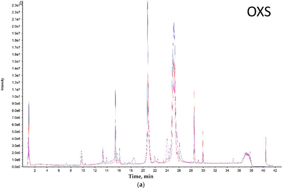
<p>Combined liquid chromatography (LC) chromatograms of specimens from the (<b>a</b>) OXS – Olkola extra small group, (<b>b</b>) OS – Olkola small group, (<b>c</b>) OM – Olkola medium group, and (<b>d</b>) OL – Olkola large group. Each colour represent a different specimen. Vertical axis is Intensity and horizontal axis is Time (minutes).</p>
Full article ">Figure 1 Cont.
<p>Combined liquid chromatography (LC) chromatograms of specimens from the (<b>a</b>) OXS – Olkola extra small group, (<b>b</b>) OS – Olkola small group, (<b>c</b>) OM – Olkola medium group, and (<b>d</b>) OL – Olkola large group. Each colour represent a different specimen. Vertical axis is Intensity and horizontal axis is Time (minutes).</p>
Full article ">Figure 2
<p>Principal component analysis and discriminant analysis (PCA-DA) analysis of <span class="html-italic">Phlogius crassipes</span> population from Olkola Aboriginal land. Tarantulas are grouped according to cephalothorax size. Each dot represents a specimen.</p>
Full article ">Figure 3
<p>Left. 1D SDS PAGE gel of representatives from a population of <span class="html-italic">Phlogius crassipes</span>. Left lane is molecular marker (protein ladder) followed by OL, OM, OS and OXS specimens, respectively. Right. Clustering analyses with Rho similarity and 10,000 bootstrap values from one-dimensional gel electrophoresis of <span class="html-italic">P. crassipes</span> individuals of four different sizes.</p>
Full article ">Figure 4
<p>Simple Correspondence Analysis of representatives of group size from a population of <span class="html-italic">P. crassipes</span>. Each number corresponds to a single venom compound identified by Protein Pilot. Axes correspond to the two dimensions created by the analysis.</p>
Full article ">

2016

Jump to: 2020, 2018, 2017, 2015, 2014, 2013

1760 KiB  
Article
Exon Shuffling and Origin of Scorpion Venom Biodiversity
by Xueli Wang, Bin Gao and Shunyi Zhu
Toxins 2017, 9(1), 10; https://doi.org/10.3390/toxins9010010 - 26 Dec 2016
Cited by 15 | Viewed by 7911
Abstract
Scorpion venom is a complex combinatorial library of peptides and proteins with multiple biological functions. A combination of transcriptomic and proteomic techniques has revealed its enormous molecular diversity, as identified by the presence of a large number of ion channel-targeted neurotoxins with different [...] Read more.
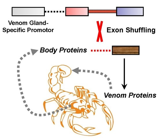
Scorpion venom is a complex combinatorial library of peptides and proteins with multiple biological functions. A combination of transcriptomic and proteomic techniques has revealed its enormous molecular diversity, as identified by the presence of a large number of ion channel-targeted neurotoxins with different folds, membrane-active antimicrobial peptides, proteases, and protease inhibitors. Although the biodiversity of scorpion venom has long been known, how it arises remains unsolved. In this work, we analyzed the exon-intron structures of an array of scorpion venom protein-encoding genes and unexpectedly found that nearly all of these genes possess a phase-1 intron (one intron located between the first and second nucleotides of a codon) near the cleavage site of a signal sequence despite their mature peptides remarkably differ. This observation matches a theory of exon shuffling in the origin of new genes and suggests that recruitment of different folds into scorpion venom might be achieved via shuffling between body protein-coding genes and ancestral venom gland-specific genes that presumably contributed tissue-specific regulatory elements and secretory signal sequences. Full article
Show Figures

Graphical abstract

Graphical abstract
Full article ">Figure 1
<p>The fold diversity of scorpion venom components. (<b>A</b>) Representative structure of three different types of peptides: MMTX (PDB: 2RTZ) (CSαβ fold), λ-MeuTx-1 (ICK fold) [<a href="#B7-toxins-09-00010" class="html-bibr">7</a>], and the α-helical Meucin-24 (PDB: 2KFE); (<b>B</b>) Representative structures of scorpion venom-derived proteases and protease inhibitors: The chymotrypsin-like protease MmChTP whose structure was modelled on SWISS-MODEL (<a href="http://www.expasy.org" target="_blank">www.expasy.org</a>) using the template of a mannose-binding lectin-associated serine proteinase-3 (PDB: 4KKD); the Kunitz-type protease inhibitor LmKTT-1a (PDB: 2M01).</p>
Full article ">Figure 2
<p>Representative gene structures of scorpion venom-derived neurotoxins and antimicrobial peptides. UTR, untranslated region; SP, signal peptide; MP, mature peptide; PP, propeptide. Functional classes: KCT, potassium channel toxin; SCT, sodium channel toxin; CCT, chloride channel toxin; DFN, defensin; RyR, ryanodine receptor; AMP, antimicrobial peptide. MM, <span class="html-italic">Mesobuthus martensii</span>; ME, <span class="html-italic">M. eupeus</span>; CE, <span class="html-italic">Centruroides exilicauda</span>; and OC, <span class="html-italic">Opistophthalmus carinatus</span>.</p>
Full article ">Figure 3
<p>Representative gene structures of scorpion venom-derived proteases (MmChTP and CsEChTP) and protease inhibitors (MmPI-1, MmPI-2a, MmPI-2b, and CsEPI-1). MM, <span class="html-italic">M. martensii</span>; CE, <span class="html-italic">C. exilicauda</span>.</p>
Full article ">Figure 4
<p>The hypothetical evolutionary model for the origin of scorpion venom biodiversity. Exon shuffling is proposed as a major evolutionary mechanism mediating the origin of venom proteins from ancestral body proteins, in which a venom gland-specific ancestral gene is considered as a donor providing two necessary elements for venom gland-specific expression: a promoter and a secretory signal.</p>
Full article ">
3135 KiB  
Article
Evolution of the Cytolytic Pore-Forming Proteins (Actinoporins) in Sea Anemones
by Jason Macrander and Marymegan Daly
Toxins 2016, 8(12), 368; https://doi.org/10.3390/toxins8120368 - 8 Dec 2016
Cited by 35 | Viewed by 6775
Abstract
Sea anemones (Cnidaria, Anthozoa, and Actiniaria) use toxic peptides to incapacitate and immobilize prey and to deter potential predators. Their toxin arsenal is complex, targeting a variety of functionally important protein complexes and macromolecules involved in cellular homeostasis. Among these, actinoporins are one [...] Read more.
Sea anemones (Cnidaria, Anthozoa, and Actiniaria) use toxic peptides to incapacitate and immobilize prey and to deter potential predators. Their toxin arsenal is complex, targeting a variety of functionally important protein complexes and macromolecules involved in cellular homeostasis. Among these, actinoporins are one of the better characterized toxins; these venom proteins form a pore in cellular membranes containing sphingomyelin. We used a combined bioinformatic and phylogenetic approach to investigate how actinoporins have evolved across three superfamilies of sea anemones (Actinioidea, Metridioidea, and Actinostoloidea). Our analysis identified 90 candidate actinoporins across 20 species. We also found clusters of six actinoporin-like genes in five species of sea anemone (Nematostella vectensis, Stomphia coccinea, Epiactis japonica, Heteractis crispa, and Diadumene leucolena); these actinoporin-like sequences resembled actinoporins but have a higher sequence similarity with toxins from fungi, cone snails, and Hydra. Comparative analysis of the candidate actinoporins highlighted variable and conserved regions within actinoporins that may pertain to functional variation. Although multiple residues are involved in initiating sphingomyelin recognition and membrane binding, there is a high rate of replacement for a specific tryptophan with leucine (W112L) and other hydrophobic residues. Residues thought to be involved with oligomerization were variable, while those forming the phosphocholine (POC) binding site and the N-terminal region involved with cell membrane penetration were highly conserved. Full article
Show Figures

Figure 1

Figure 1
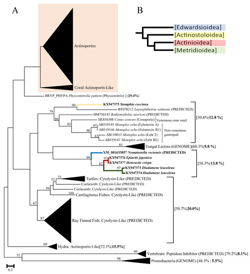
<p>(<b>A</b>) Maximum Likelihood tree of actinoporins and actinoporin-like proteins produced in FastTree2. Numbers on branches represent bootstrap values of 1000 replicates. Bootstrapping values greater than 50 are shown at the nodes. Branch labels of clustered gene groups represent lineage common names followed by percent identity (identical amino acid residues) within gene cluster and percent identity when compared to actinoporins in bold. Labeled individual branches show GenBank accession followed by species, and protein name (if applicable). Labels denoted with PREDICTED or GENOME indicate that they were derived bioinformatically in GenBank and are not validated proteins. Branch labels with Genbank accession and species for sea anemones are indicated in bold. The colored box indicates which actinoporin sequences were used in subsequent analyses; (<b>B</b>) Phylogenetic tree with branch colors depicting superfamily associations (Blue: Edwardsioidea, Yellow: Actinostoloidea, Red: Actinioidea, Green: Metridioidea) [<a href="#B42-toxins-08-00368" class="html-bibr">42</a>].</p>
Full article ">Figure 2
<p>(<b>A</b>) Functionally important residues identified on EqII [<a href="#B43-toxins-08-00368" class="html-bibr">43</a>]. Functional sites are as follows: (B) site of bend when N-terminus comes into contact with the cell membrane, (POC) residues involved with the POC binding site, (O) residues involved with oligomerization, (S) key sphingomyelin binding site. Colors are used to aid in determining the orientation between the two views shown; (<b>B</b>) Characterization of amino acid variation for the different gene clusters (Clade 1, Clade 2M, Clade 2A) identified in our analysis. SeqLogo graphs for residues that have been identified previously as functionally important above the alignment with the size of each amino acid residue representing the frequency in which these residues occurred in the alignment. Numbers along the bottom correspond to positions of specific amino acid residues in EqII; (<b>C</b>) Maximum Likelihood actinoporin gene tree produced in FastTree2. Colored branches depict superfamily associations (see <a href="#toxins-08-00368-f001" class="html-fig">Figure 1</a>, Yellow: Actinostoloidea, Red: Actinioidea, Green: Metridioidea). Bootstrapping values greater than 50 are shown at the nodes. Branch labels include GenBank ID (when applicable) and the species from which the toxin gene was derived. Bold labels indicate that the mature protein sequence was recovered. Sequences derived from genomic data are indicated with “G” following species in sequence IDs. The superfamily association for <span class="html-italic">Actineria villosa</span> may be incorrect and is noted with an asterisk.</p>
Full article ">Figure 3
<p>Edmundson wheel projections were determined in HeliQuest. Associated Sequence IDs are shown below the species name.</p>
Full article ">Figure 3 Cont.
<p>Edmundson wheel projections were determined in HeliQuest. Associated Sequence IDs are shown below the species name.</p>
Full article ">
1044 KiB  
Article
A Tricky Trait: Applying the Fruits of the “Function Debate” in the Philosophy of Biology to the “Venom Debate” in the Science of Toxinology
by Timothy N. W. Jackson and Bryan G. Fry
Toxins 2016, 8(9), 263; https://doi.org/10.3390/toxins8090263 - 7 Sep 2016
Cited by 30 | Viewed by 7811
Abstract
The “function debate” in the philosophy of biology and the “venom debate” in the science of toxinology are conceptually related. Venom systems are complex multifunctional traits that have evolved independently numerous times throughout the animal kingdom. No single concept of function, amongst those [...] Read more.
The “function debate” in the philosophy of biology and the “venom debate” in the science of toxinology are conceptually related. Venom systems are complex multifunctional traits that have evolved independently numerous times throughout the animal kingdom. No single concept of function, amongst those popularly defended, appears adequate to describe these systems in all their evolutionary contexts and extant variations. As such, a pluralistic view of function, previously defended by some philosophers of biology, is most appropriate. Venom systems, like many other functional traits, exist in nature as points on a continuum and the boundaries between “venomous” and “non-venomous” species may not always be clearly defined. This paper includes a brief overview of the concept of function, followed by in-depth discussion of its application to venom systems. A sound understanding of function may aid in moving the venom debate forward. Similarly, consideration of a complex functional trait such as venom may be of interest to philosophers of biology. Full article
Show Figures

Figure 1

Figure 1
<p>Flow chart illustrating basic application of <span class="html-italic">evolutionary</span> theories of function to properties of traits.</p>
Full article ">Figure 2
<p>Flow chart illustrating functional analysis of <span class="html-italic">exophysiological</span> secretion in order to determine its classification as venom, poison, digestive aid, antimicrobial, or other. An exophysiological secretion is a secretion containing molecules that have evolved to be physiologically active outside the body of the producing organism (e.g., in the body of another organism). Note that this functional analysis is intended as an example only and is not exhaustive.</p>
Full article ">
2305 KiB  
Article
Canopy Venom: Proteomic Comparison among New World Arboreal Pit-Viper Venoms
by Jordan Debono, Chip Cochran, Sanjaya Kuruppu, Amanda Nouwens, Niwanthi W. Rajapakse, Minami Kawasaki, Kelly Wood, James Dobson, Kate Baumann, Mahdokht Jouiaei, Timothy N. W. Jackson, Ivan Koludarov, Dolyce Low, Syed A. Ali, A. Ian Smith, Andrew Barnes and Bryan G. Fry
Toxins 2016, 8(7), 210; https://doi.org/10.3390/toxins8070210 - 8 Jul 2016
Cited by 7 | Viewed by 7099
Abstract
Central and South American pitvipers, belonging to the genera Bothrops and Bothriechis, have independently evolved arboreal tendencies. Little is known regarding the composition and activity of their venoms. In order to close this knowledge gap, venom proteomics and toxin activity of species [...] Read more.
Central and South American pitvipers, belonging to the genera Bothrops and Bothriechis, have independently evolved arboreal tendencies. Little is known regarding the composition and activity of their venoms. In order to close this knowledge gap, venom proteomics and toxin activity of species of Bothriechis, and Bothrops (including Bothriopsis) were investigated through established analytical methods. A combination of proteomics and bioactivity techniques was used to demonstrate a similar diversification of venom composition between large and small species within Bothriechis and Bothriopsis. Increasing our understanding of the evolution of complex venom cocktails may facilitate future biodiscoveries. Full article
Show Figures

Figure 1

Figure 1
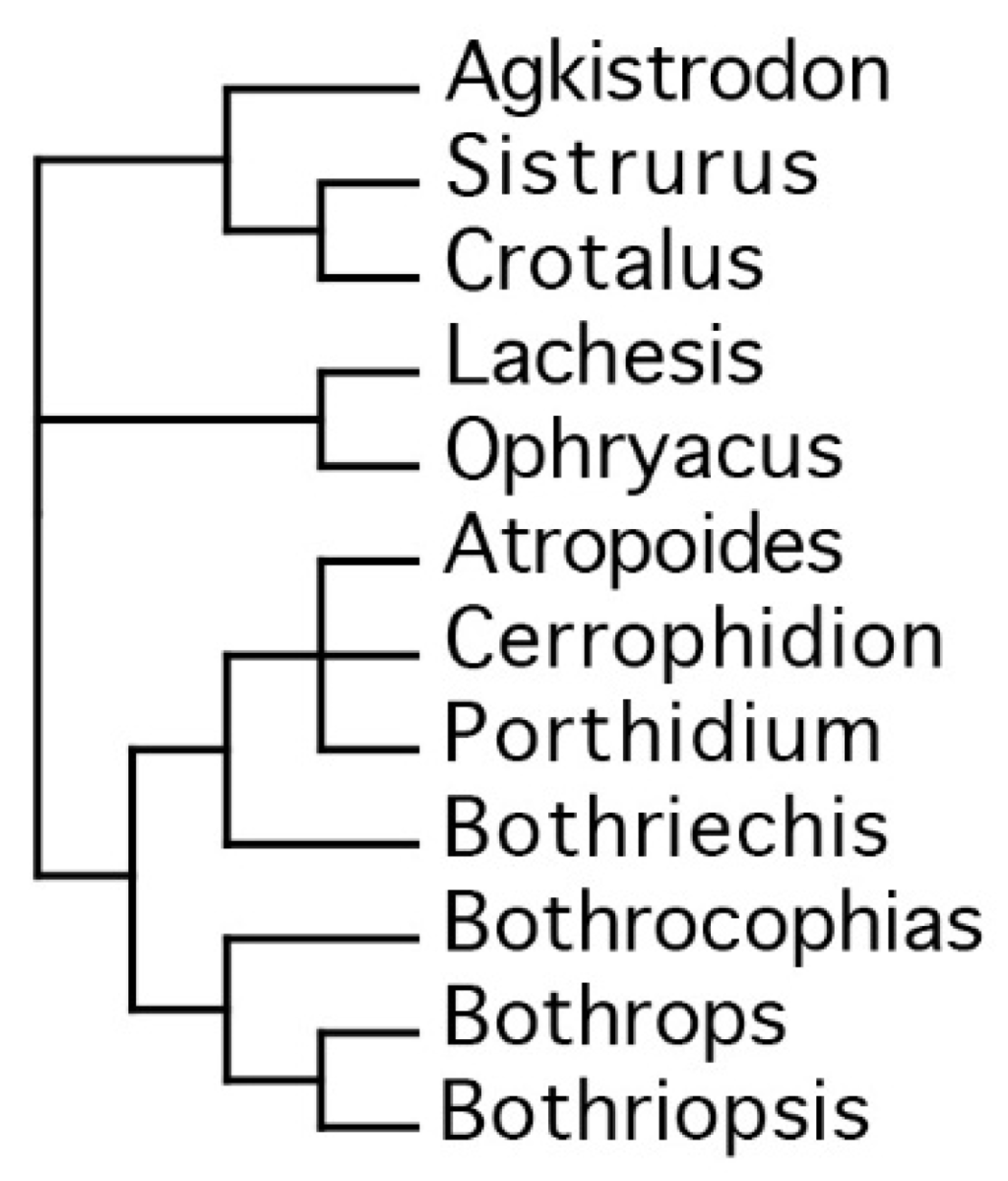
<p>Taxonomical relationships of American pitvipers, based on Castoe 2006 [<a href="#B65-toxins-08-00210" class="html-bibr">65</a>].</p>
Full article ">Figure 2
<p>1D SDS page. MW = molecular weight marker; aur = <span class="html-italic">Bothriechis aurifer</span>; lat = <span class="html-italic">Bothriechis lateralis</span>; mar = <span class="html-italic">Bothriechis marchi</span>; sch = <span class="html-italic">Bothriechis schlegelli</span>; bil = <span class="html-italic">Bothrops bilineata</span>; tan = <span class="html-italic">Bothrops taeniata</span>; asp = <span class="html-italic">Bothrops asper</span>; neu = <span class="html-italic">Bothrops neuwiedi bolivianus</span>. Annotation indicates the dominant type in a region. However, other toxin types may also be present (see <a href="#app1-toxins-08-00210" class="html-app">Supplementary File 1</a> for full annotation).</p>
Full article ">Figure 3
<p>2D SDS page analysis of (<b>A</b>) <span class="html-italic">Bothriechis aurifer</span>; (<b>B</b>) <span class="html-italic">Bothriechis lateralis</span>; (<b>C</b>) <span class="html-italic">Bothriechis marchi</span>; (<b>D</b>) <span class="html-italic">Bothriechis schlegelli</span>; (<b>E</b>) <span class="html-italic">Bothrops bilineata</span>; (<b>F</b>) <span class="html-italic">Bothrops taeniata</span>; (<b>G</b>) <span class="html-italic">Bothrops asper</span> and (<b>H</b>) <span class="html-italic">Bothrops neuwiedi bolivianus</span>. pI range is 3–10 (left to right) and molecular weight markers are as for <a href="#toxins-08-00210-f002" class="html-fig">Figure 2</a>.</p>
Full article ">Figure 4
<p>Metalloprotease activity of venom—Metalloprotease activity of venom (10 ng/µL) was measured based on its ability to cleave a fluorogenic peptide substrate (Mca-PLGL-Dpa-AR-NH<sub>2</sub>, 10 µM final). aur = <span class="html-italic">Bothriechis aurifer;</span> lat = <span class="html-italic">Bothriechis lateralis;</span> mar = <span class="html-italic">Bothriechis marchi;</span> sch = <span class="html-italic">Bothriechis schlegelii</span>; bil = <span class="html-italic">Bothrops bilineata;</span> tan = <span class="html-italic">Bothrops taeniata;</span> asp = <span class="html-italic">Bothrops asper;</span> neu = <span class="html-italic">Bothrops neuwiedi bolivianus.</span></p>
Full article ">Figure 5
<p>Crude venom zymography gel analysis using casein (<b>A</b>) and gelatin (<b>B</b>) as protein substrates. MW = molecular weight marker; aur = <span class="html-italic">Bothriechis aurifer;</span> lat = <span class="html-italic">Bothriechis lateralis;</span> mar = <span class="html-italic">Bothriechis marchi;</span> sch = <span class="html-italic">Bothriechis schlegelii</span>; bil = <span class="html-italic">Bothrops bilineata;</span> tan = <span class="html-italic">Bothrops taeniata;</span> asp = <span class="html-italic">Bothrops asper;</span> neu = <span class="html-italic">Bothrops neuwiedi bolivianus.</span></p>
Full article ">Figure 6
<p>Phospholipase A<sub>2</sub> enzymatic activity profiling measured by means of absorbance over time at 1 µg for (<b>A</b>) <span class="html-italic">Bothriechis</span> species; (<b>B</b>) arboreal <span class="html-italic">Bothrops</span> species and (<b>C</b>) terrestrial <span class="html-italic">Bothrops</span> species.</p>
Full article ">
223 KiB  
Article
Tempo and Mode of the Evolution of Venom and Poison in Tetrapods
by Richard J. Harris and Kevin Arbuckle
Toxins 2016, 8(7), 193; https://doi.org/10.3390/toxins8070193 - 23 Jun 2016
Cited by 24 | Viewed by 7775
Abstract
Toxic weaponry in the form of venom and poison has evolved in most groups of animals, including all four major lineages of tetrapods. Moreover, the evolution of such traits has been linked to several key aspects of the biology of toxic animals including [...] Read more.
Toxic weaponry in the form of venom and poison has evolved in most groups of animals, including all four major lineages of tetrapods. Moreover, the evolution of such traits has been linked to several key aspects of the biology of toxic animals including life-history and diversification. Despite this, attempts to investigate the macroevolutionary patterns underlying such weaponry are lacking. In this study we analyse patterns of venom and poison evolution across reptiles, amphibians, mammals, and birds using a suite of phylogenetic comparative methods. We find that each major lineage has a characteristic pattern of trait evolution, but mammals and reptiles evolve under a surprisingly similar regime, whilst that of amphibians appears to be particularly distinct and highly contrasting compared to other groups. Our results also suggest that the mechanism of toxin acquisition may be an important distinction in such evolutionary patterns; the evolution of biosynthesis is far less dynamic than that of sequestration of toxins from the diet. Finally, contrary to the situation in amphibians, other tetrapod groups show an association between the evolution of toxic weaponry and higher diversification rates. Taken together, our study provides the first broad-scale analysis of macroevolutionary patterns of venom and poison throughout tetrapods. Full article
Show Figures

Graphical abstract

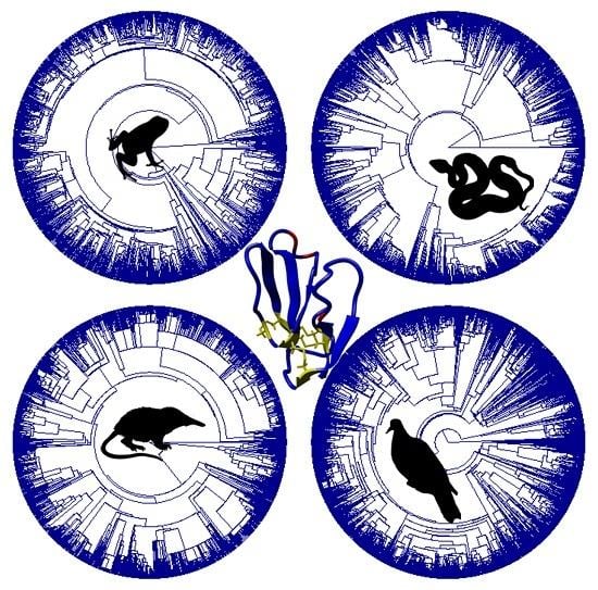
Graphical abstract
Full article ">
2296 KiB  
Article
Is Hybridization a Source of Adaptive Venom Variation in Rattlesnakes? A Test, Using a Crotalus scutulatus × viridis Hybrid Zone in Southwestern New Mexico
by Giulia Zancolli, Timothy G. Baker, Axel Barlow, Rebecca K. Bradley, Juan J. Calvete, Kimberley C. Carter, Kaylah De Jager, John Benjamin Owens, Jenny Forrester Price, Libia Sanz, Amy Scholes-Higham, Liam Shier, Liam Wood, Catharine E. Wüster and Wolfgang Wüster
Toxins 2016, 8(6), 188; https://doi.org/10.3390/toxins8060188 - 16 Jun 2016
Cited by 30 | Viewed by 11376
Abstract
Venomous snakes often display extensive variation in venom composition both between and within species. However, the mechanisms underlying the distribution of different toxins and venom types among populations and taxa remain insufficiently known. Rattlesnakes (Crotalus, Sistrurus) display extreme inter- and [...] Read more.
Venomous snakes often display extensive variation in venom composition both between and within species. However, the mechanisms underlying the distribution of different toxins and venom types among populations and taxa remain insufficiently known. Rattlesnakes (Crotalus, Sistrurus) display extreme inter- and intraspecific variation in venom composition, centered particularly on the presence or absence of presynaptically neurotoxic phospholipases A2 such as Mojave toxin (MTX). Interspecific hybridization has been invoked as a mechanism to explain the distribution of these toxins across rattlesnakes, with the implicit assumption that they are adaptively advantageous. Here, we test the potential of adaptive hybridization as a mechanism for venom evolution by assessing the distribution of genes encoding the acidic and basic subunits of Mojave toxin across a hybrid zone between MTX-positive Crotalus scutulatus and MTX-negative C. viridis in southwestern New Mexico, USA. Analyses of morphology, mitochondrial and single copy-nuclear genes document extensive admixture within a narrow hybrid zone. The genes encoding the two MTX subunits are strictly linked, and found in most hybrids and backcrossed individuals, but not in C. viridis away from the hybrid zone. Presence of the genes is invariably associated with presence of the corresponding toxin in the venom. We conclude that introgression of highly lethal neurotoxins through hybridization is not necessarily favored by natural selection in rattlesnakes, and that even extensive hybridization may not lead to introgression of these genes into another species. Full article
Show Figures

Graphical abstract

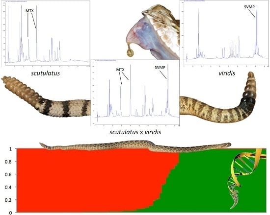
Graphical abstract
Full article ">Figure 1
<p>Ordination of specimens of <span class="html-italic">C. scutulatus</span>, <span class="html-italic">C. atrox</span>, <span class="html-italic">C. viridis</span> and putative hybrids along the first two axes of a principal components analysis of nine morphological characters. All specimens from the eastern slope of the Peloncillo Mountains in southwestern New Mexico are labeled as hybrids irrespective of morphological or genetic profile. The first and second principal components represent 41.5% and 23.7% of the total variance, respectively.</p>
Full article ">Figure 2
<p>Neighbor-joining tree of individual ND4 sequences. Tip labels with yellow background indicate specimens from the putative hybrid zone in SW New Mexico.</p>
Full article ">Figure 3
<p>(<b>A</b>) Bayesian population clustering of individuals of <span class="html-italic">C. atrox</span>, <span class="html-italic">C. scutulatus</span>, <span class="html-italic">C. viridis</span> and putative hybrids based on allele frequencies of four single copy nuclear genes. Above the Structure clustering, rows of boxes indicate mtDNA haplotype affinities (same colors as nuclear structuring), and the presence (black) or absence (grey) of the genes coding for the basic and acidic subunits of Mojave toxin (MTX), and above the confirmed presence or absence of the corresponding proteins in the venom. White spaces indicate absence of data. Black arrows indicate specimens morphologically intermediate between <span class="html-italic">C. scutulatus</span> and <span class="html-italic">C. atrox</span>. (<b>B</b>) Equivalent analysis excluding <span class="html-italic">C. atrox</span> to emphasize hybrid zone between <span class="html-italic">C. scutulatus</span> and <span class="html-italic">C. viridis</span>.</p>
Full article ">Figure 4
<p>Sampling localities, hybrid status and MTX status of individuals of <span class="html-italic">C. scutulatus</span>, <span class="html-italic">C. viridis</span> and their hybrids. Diamonds indicate MTX+ve, circles MTX−ve individuals, the degree of shading of the symbols indicates the proportion of the genome attributed to <span class="html-italic">C. scutulatus</span> in the Structure analysis.</p>
Full article ">Figure 5
<p>Examples of reverse-phase high performance liquid chromatography (RP-HPLC) chromatograms from different venoms included in the study. A. <span class="html-italic">Crotalus scutulatus</span> type II venom with MTX but lacking snake venom metalloproteinases (SVMPs); sample 4311, nr. Rodeo, Hidalgo Co., NM. B. <span class="html-italic">Crotalus scutulatus</span> × <span class="html-italic">viridis</span>, hybrid containing MTX and SVMPs; sample 4687, nr. Cotton City, Hidalgo Co., NM. C. <span class="html-italic">Crotalus viridis</span>, typical venom lacking MTX but containing SVMPs; sample 4590, nr. Animas, Hidalgo Co., NM. D. <span class="html-italic">Crotalus atrox</span>, typical venom lacking MTX but containing SVMPs; sample 4594, nr. Benson, Cochise Co., AZ.</p>
Full article ">

2015

Jump to: 2020, 2018, 2017, 2016, 2014, 2013

701 KiB  
Review
Cabinet of Curiosities: Venom Systems and Their Ecological Function in Mammals, with a Focus on Primates
by Johanna E. Rode-Margono and K. Anne-Isola Nekaris
Toxins 2015, 7(7), 2639-2658; https://doi.org/10.3390/toxins7072639 - 17 Jul 2015
Cited by 32 | Viewed by 16824
Abstract
Venom delivery systems (VDS) are common in the animal kingdom, but rare amongst mammals. New definitions of venom allow us to reconsider its diversity amongst mammals by reviewing the VDS of Chiroptera, Eulipotyphla, Monotremata, and Primates. All orders use modified anterior dentition as [...] Read more.
Venom delivery systems (VDS) are common in the animal kingdom, but rare amongst mammals. New definitions of venom allow us to reconsider its diversity amongst mammals by reviewing the VDS of Chiroptera, Eulipotyphla, Monotremata, and Primates. All orders use modified anterior dentition as the venom delivery apparatus, except Monotremata, which possesses a crural system. The venom gland in most taxa is a modified submaxillary salivary gland. In Primates, the saliva is activated when combined with brachial gland exudate. In Monotremata, the crural spur contains the venom duct. Venom functions include feeding, intraspecific competition, anti-predator defense and parasite defense. Including mammals in discussion of venom evolution could prove vital in our understanding protein functioning in mammals and provide a new avenue for biomedical and therapeutic applications and drug discovery. Full article
Show Figures

Graphical abstract

Graphical abstract
Full article ">Figure 1
<p>Venom system of vampire bats. Common vampire bat <span class="html-italic">Desmodus rotundus</span> (<b>a</b>); with specialized tongue as indicated by the arrow (<b>b</b>); and teeth (<b>c</b>) Illustrations: Kathleen Reinhardt.</p>
Full article ">Figure 2
<p>European water shrew <span class="html-italic">Neomys fodiens</span> (<b>a</b>); with concave incisor surfaces (as indicated by the arrow) that help with flow and injection of venom (<b>b</b>). Illustrations: Kathleen Reinhardt.</p>
Full article ">Figure 3
<p>Venom system of solenodons. Hispaniolan solenodon <span class="html-italic">Solenodon paradoxus</span> (<b>a</b>); with deeply grooved lower canines (as indicated by the arrow) that aid in flow and injection of venom (<b>b</b>). Illustrations: Kathleen Reinhardt.</p>
Full article ">Figure 4
<p>Venom system of the platypus. Platypus <span class="html-italic">Ornithorhynchus anatinus</span> (<b>a</b>); with crural spur as indicated by the arrow (<b>b</b>). Illustrations: Kathleen Reinhardt.</p>
Full article ">Figure 5
<p>Venom system of slow lorises. Javan slow loris <span class="html-italic">Nycticebus javanicus</span> showing warning coloration of face (<b>a</b>); Javan slow loris displaying defense position (<b>b</b>); brachial gland as indicated by the arrow (<b>c</b>); tooth comb as indicated by the arrow (<b>d</b>); Illustrations: Kathleen Reinhardt.</p>
Full article ">
634 KiB  
Review
Facing Hymenoptera Venom Allergy: From Natural to Recombinant Allergens
by Amilcar Perez-Riverol, Débora Lais Justo-Jacomini, Ricardo De Lima Zollner and Márcia Regina Brochetto-Braga
Toxins 2015, 7(7), 2551-2570; https://doi.org/10.3390/toxins7072551 - 9 Jul 2015
Cited by 27 | Viewed by 8031
Abstract
Along with food and drug allergic reactions, a Hymenoptera insect Sting (Apoidea, Vespidae, Formicidae) is one of the most common causes of anaphylaxis worldwide. Diagnoses of Hymenoptera venom allergy (HVA) and specific immunotherapy (SIT) have been based on the use of crude venom [...] Read more.
Along with food and drug allergic reactions, a Hymenoptera insect Sting (Apoidea, Vespidae, Formicidae) is one of the most common causes of anaphylaxis worldwide. Diagnoses of Hymenoptera venom allergy (HVA) and specific immunotherapy (SIT) have been based on the use of crude venom extracts. However, the incidence of cross-reactivity and low levels of sensibility during diagnosis, as well as the occurrence of nonspecific sensitization and undesired side effects during SIT, encourage the search for novel allergenic materials. Recombinant allergens are an interesting approach to improve allergy diagnosis and SIT because they circumvent major problems associated with the use of crude venom. Production of recombinant allergens depends on the profound molecular characterization of the natural counterpart by combining some “omics” approaches with high-throughput screening techniques and the selection of an appropriate system for heterologous expression. To date, several clinically relevant allergens and novel venom toxins have been identified, cloned and characterized, enabling a better understanding of the whole allergenic and envenoming processes. Here, we review recent findings on identification, molecular characterization and recombinant expression of Hymenoptera venom allergens and on the evaluation of these heterologous proteins as valuable tools for tackling remaining pitfalls on HVA diagnosis and immunotherapy. Full article

2014

Jump to: 2020, 2018, 2017, 2016, 2015, 2013

2598 KiB  
Review
Quo Vadis Venomics? A Roadmap to Neglected Venomous Invertebrates
by Bjoern Marcus Von Reumont, Lahcen I. Campbell and Ronald A. Jenner
Toxins 2014, 6(12), 3488-3551; https://doi.org/10.3390/toxins6123488 - 19 Dec 2014
Cited by 86 | Viewed by 22729
Abstract
Venomics research is being revolutionized by the increased use of sensitive -omics techniques to identify venom toxins and their transcripts in both well studied and neglected venomous taxa. The study of neglected venomous taxa is necessary both for understanding the full diversity of [...] Read more.
Venomics research is being revolutionized by the increased use of sensitive -omics techniques to identify venom toxins and their transcripts in both well studied and neglected venomous taxa. The study of neglected venomous taxa is necessary both for understanding the full diversity of venom systems that have evolved in the animal kingdom, and to robustly answer fundamental questions about the biology and evolution of venoms without the distorting effect that can result from the current bias introduced by some heavily studied taxa. In this review we draw the outlines of a roadmap into the diversity of poorly studied and understood venomous and putatively venomous invertebrates, which together represent tens of thousands of unique venoms. The main groups we discuss are crustaceans, flies, centipedes, non-spider and non-scorpion arachnids, annelids, molluscs, platyhelminths, nemerteans, and echinoderms. We review what is known about the morphology of the venom systems in these groups, the composition of their venoms, and the bioactivities of the venoms to provide researchers with an entry into a large and scattered literature. We conclude with a short discussion of some important methodological aspects that have come to light with the recent use of new -omics techniques in the study of venoms. Full article
Show Figures

Figure 1

Figure 1
<p>The first venomous crustacean, <span class="html-italic">Xibalbanus tulumensis</span>. (<b>A</b>) The cephalon and part of the trunk is shown from the ventral side. TrA = trunk appendages, Vg = venom gland, Mxp = maxilliped, Max 2 = maxilla 2, Max1 = maxilla 1 (= maxillule), Ant = antenna (antennula); (<b>B</b>) An individual (marked in red circle) is caught in the Mexican anchialine cave system “Cenote Crustacea” by BMvR; (<b>C</b>) Habitus of a specimen, showing its remarkably convergent bodyplan to centipedes with homonomously segmented trunk and similar trunk appendages (TrA).</p>
Full article ">Figure 2
<p>Synchrotron-based computer tomographic reconstruction of the cephalothorax and the venom delivery system of the remipede <span class="html-italic">Xibalbanus tulumensis</span> in lateral view (anterior to the left), (<b>A</b>), and in ventral view (<b>B</b>), and the muscle system that facilitates venom injection by the maxillule (<b>C</b>); (<b>D</b>) shows the composition of the cocktail of toxin gene transcripts expressed in the venom glands. Abbreviations: 4 seg = 4th segments of maxillule, ab = abductor muscles, ad = adductor muscles br = brain, cep = cephalothorax, phx = pharynx, mxu = maxillula, t = tegument, vnc = ventral nerv cord, gl = venom gland, dc = venom duct, rv = venom reservoir, vm = ventral apodemal muscle.</p>
Full article ">Figure 3
<p>Other putative venomous crustaceans. (<b>A</b>) Branchiuran carp louse <span class="html-italic">Argulus foliacaetus</span> from ventral side; (<b>B</b>) <span class="html-italic">Argulus</span> mouthparts from ventral side. Thp 1–4 = thoracopods 1–4, Max 1 = maxilla 1 (maxillule) (with last segment modified to sucker disc), Max 2 = maxilla 2, Mc = mouth cone, Ps = Preoral spine (slightly disarranged by ethanol preservation); (<b>C</b>) Male skeleton shrimp <span class="html-italic">Caprella scaura</span> with pleiopods removed. Inset shows gnathopod 2 appendages, which bear the poison tooth. Gnp = gnathopod, Pot = poison tooth (<b>D</b>) Female sea lice or siphonostomatoid copepods. Collection reference numbers for specimens of the Natural History Museum London: <span class="html-italic">Caprella Scaura</span>, 1902, male, Inland sea, Japan (Amphipods, Caprellidea: NHMUK 1902.12.12.6/7). <span class="html-italic">Caligus rogercresseyi</span>, 2000, female, Puerto Montt Chile, Host <span class="html-italic">Eleginops maclovinus</span>, J. Carvaja: (Copepods, Siphonostomatoidae: 2000.1258-126). Specimens were photographed with a Nikon D200, Sigma 150 mm EX-APO Macro-lens and ring-flashgun units.</p>
Full article ">Figure 4
<p>Examples of fly groups that have neglected, likely venomous species. (<b>A</b>) Robber flies (Asilidae): <span class="html-italic">Promachus leoninus</span> specimens and larva of a British robber fly; (<b>B</b>) Marsh flies (Sciomyzidae): <span class="html-italic">Tetanocera elata</span>; (<b>C</b>) Horse flies (Tabanidae): <span class="html-italic">Tabanus trigonus</span>, one of the species of which larvae pose a risk to rice workers in Japan. Collection reference numbers for specimens of the Natural History Museum London: <span class="html-italic">Promachus leoninus</span>, Turkey, Nurdagi Gecidi, 1960 (DIP3404, B35 193, Bactria, BMNH(E) 1237803). <span class="html-italic">Tetanocera elata</span>, United Kingdom, 1971, (DIP957, C57, 8, BMNH(E)1237801). <span class="html-italic">Tabanus trigonus</span> 1972, Japan, Kasumigaura Ibaraki (DIP3026B5, 189, Tabanidae, BMNH(E) 1237802).</p>
Full article ">Figure 5
<p>Representative species of neglected but common centipede groups. (<b>A</b>) <span class="html-italic">Lithobius forficatus</span> (Lithobiomorpha); (<b>B</b>) <span class="html-italic">Cryptops</span> sp. (Scolopendromorpha); (<b>C</b>) <span class="html-italic">Alipes grandidieri</span> (Scolopendromorpha) (<b>D</b>) <span class="html-italic">Scutigera coleoptrata</span> (Scutigeromorpha).</p>
Full article ">Figure 6
<p>Pseudoscorpions. (<b>A</b>) Unidentified pseudoscorpion from New Zealand guarding its eggs; (<b>B</b>) Unidentified pseudoscorpion from New Zealand. Copyright for both photos is with Gonzalo Giribet, and are reproduced with his permission.</p>
Full article ">Figure 7
<p>The venom glands in the palpal hand of: (<b>A</b>) <span class="html-italic">Cordylochernes macrochelatus</span>; (<b>B</b>) <span class="html-italic">Shravana laminata</span>; (<b>C</b>) <span class="html-italic">Neobisium flexifemoratum</span>. ff = fixed finger; mf = moveable finger; vg = venom gland; vt = venom tooth. Figure redrawn and modified from Weygoldt [<a href="#B130-toxins-06-03488" class="html-bibr">130</a>].</p>
Full article ">Figure 8
<p>(<b>A</b>) Anatomy and general morphology of a <span class="html-italic">Glycera</span> bloodworm; (<b>B</b>) Rendered micro-CT picture of longitudinal section through everted four jaws and proboscis (<b>left</b>), and outer view from above (<b>right</b>) of <span class="html-italic">Glycera tesselata</span>. glm = muscles associated with the venom glands; pap = parapodium; prb = proboscis; pst = prostomium; vdc = venom duct; vgl = venom gland.</p>
Full article ">Figure 9
<p>Transcriptome profile of toxin genes expressed in the venom glands of <span class="html-italic">Glycera dibranchiata</span>. Pie chart shows the contig diversity of the 20 different toxins expressed in the most deeply sequenced species <span class="html-italic">Glycera dibranchiata</span>. Relative contig diversity is expressed as percentages followed by total numbers of contigs in parentheses. See [<a href="#B155-toxins-06-03488" class="html-bibr">155</a>] for full details.</p>
Full article ">Figure 10
<p>General body morphology of scale-worm polychaetes. Individual shown is an unidentified polynoid species. Copyright for this picture resides with Helena Wiklund, and is reproduced with her permission.</p>
Full article ">Figure 11
<p>Predating nemerteans. (<b>A</b>) Heteronemertean species <span class="html-italic">Ramphogordius sanguineus</span> feeding on the polychaete <span class="html-italic">Alitta succinea</span>; (<b>B</b>) Hoplonemertean species <span class="html-italic">Prosorhochmus nelsoni</span> feeding on the isopod <span class="html-italic">Ligia</span> sp. Copyright of both photos is with Serena Caplins, and are reproduced with her permission.</p>
Full article ">Figure 12
<p>Venom appendages of sea urchins (Echinoidea). (<b>A</b>) A secondary aboral spine of <span class="html-italic">Asthenosoma vaium,</span> showing spine tip and associated venom gland (sac) and muscle tissue; (<b>B</b>) Typical venomous globiferous pedicellaria. Pedicellaria shown is fanged with external venom glands on the valves. Distinct from “Fistulate” globiferous pedicellaria (internally located venom glands) [<a href="#B284-toxins-06-03488" class="html-bibr">284</a>]. Figure redrawn and modified from Halstead [<a href="#B274-toxins-06-03488" class="html-bibr">274</a>].</p>
Full article ">Figure 13
<p>Flowchart of transcriptomic analyses using NGS data to identify putative toxin proteins. Software used to conduct analyses is indicated in square brackets; please keep in mind that these are examples and not an exhaustive presentation of possible software.</p>
Full article ">
697 KiB  
Article
The Finding of a Group IIE Phospholipase A2 Gene in a Specified Segment of Protobothrops flavoviridis Genome and Its Possible Evolutionary Relationship to Group IIA Phospholipase A2 Genes
by Kazuaki Yamaguchi, Takahito Chijiwa, Naoki Ikeda, Hiroki Shibata, Yasuyuki Fukumaki, Naoko Oda-Ueda, Shosaku Hattori and Motonori Ohno
Toxins 2014, 6(12), 3471-3487; https://doi.org/10.3390/toxins6123471 - 18 Dec 2014
Cited by 6 | Viewed by 7194
Abstract
The genes encoding group IIE phospholipase A2, abbreviated as IIE PLA2, and its 5' and 3' flanking regions of Crotalinae snakes such as Protobothrops flavoviridis, P. tokarensis, P. elegans, and Ovophis okinavensis, were found and [...] Read more.
The genes encoding group IIE phospholipase A2, abbreviated as IIE PLA2, and its 5' and 3' flanking regions of Crotalinae snakes such as Protobothrops flavoviridis, P. tokarensis, P. elegans, and Ovophis okinavensis, were found and sequenced. The genes consisted of four exons and three introns and coded for 22 or 24 amino acid residues of the signal peptides and 134 amino acid residues of the mature proteins. These IIE PLA2s show high similarity to those from mammals and Colubridae snakes. The high expression level of IIE PLA2s in Crotalinae venom glands suggests that they should work as venomous proteins. The blast analysis indicated that the gene encoding OTUD3, which is ovarian tumor domain-containing protein 3, is located in the 3' downstream of IIE PLA2 gene. Moreover, a group IIA PLA2 gene was found in the 5' upstream of IIE PLA2 gene linked to the OTUD3 gene (OTUD3) in the P. flavoviridis genome. It became evident that the specified arrangement of IIA PLA2 gene, IIE PLA2 gene, and OTUD3 in this order is common in the genomes of humans to snakes. The present finding that the genes encoding various secretory PLA2s form a cluster in the genomes of humans to birds is closely related to the previous finding that six venom PLA2 isozyme genes are densely clustered in the so-called NIS-1 fragment of the P. flavoviridis genome. It is also suggested that venom IIA PLA2 genes may be evolutionarily derived from the IIE PLA2 gene. Full article
Show Figures

Graphical abstract

Graphical abstract
Full article ">Figure 1
<p>The schematic representation of the genome segment harboring the Crotalinae IIE PLA<sub>2</sub> gene. The nucleotide positions are numbered. Closed boxes represent open reading frames (ORFs) and open boxes untranslated regions (UTRs). Vertical bars indicate the positions of restriction enzyme sites. Arrow heads show the positions of primers.</p>
Full article ">Figure 2
<p>The aligned amino acid sequences of IIE, IIA, IA and IB PLA<sub>2</sub>s from snakes and mammals. The positions are numbered from the first residue of the signal peptides. The half-cystines are shown in shaded letters. Abbreviations: <span class="html-italic">Dt</span>, <span class="html-italic">Dispholidus typus</span>; <span class="html-italic">Hs</span>, <span class="html-italic">Homo sapiens</span>; <span class="html-italic">Lm</span>, <span class="html-italic">Leioheterodon madagascariensis</span>; <span class="html-italic">Ls</span>, <span class="html-italic">Laticauda semifasciata</span>; <span class="html-italic">Mm</span>, <span class="html-italic">Mus musculus</span>; <span class="html-italic">Oo</span>, <span class="html-italic">Ovophis okinavensis</span>; <span class="html-italic">Pe</span>, <span class="html-italic">Protobothrops elegans</span>; <span class="html-italic">Pf</span>, <span class="html-italic">P. flavoviridis</span>; and <span class="html-italic">Pt</span>, <span class="html-italic">P. tokarensis</span>. References: <span class="html-italic">Pf</span>IIEPLA<sub>2</sub> (this work); <span class="html-italic">Pt</span>IIEPLA<sub>2</sub> (this work); <span class="html-italic">Pe</span>IIEPLA<sub>2</sub> (this work); <span class="html-italic">Oo</span>IIEPLA<sub>2</sub> (this work); <span class="html-italic">Dt</span>Dis-1 (AFH66958) [<a href="#B17-toxins-06-03471" class="html-bibr">17</a>]; <span class="html-italic">Hs</span>IIEPLA<sub>2</sub> (NP_055404) [<a href="#B19-toxins-06-03471" class="html-bibr">19</a>]; <span class="html-italic">Lm</span>Lei-1 (AFH66960) [<a href="#B17-toxins-06-03471" class="html-bibr">17</a>]; <span class="html-italic">Ls</span>LsPLA2cPm09 (BAB03302) [<a href="#B32-toxins-06-03471" class="html-bibr">32</a>]; <span class="html-italic">Mm</span>IIEPLA<sub>2</sub> (NP_036174) [<a href="#B20-toxins-06-03471" class="html-bibr">20</a>]; <span class="html-italic">Pf</span>PLA-B (BAG82670) [<a href="#B13-toxins-06-03471" class="html-bibr">13</a>]; <span class="html-italic">Pf</span>PLA-N (BAG82669) [<a href="#B13-toxins-06-03471" class="html-bibr">13</a>]; <span class="html-italic">PfPLA 6</span> (BAJ84552) [<a href="#B29-toxins-06-03471" class="html-bibr">29</a>]; and <span class="html-italic">Pf</span>PancPLA<sub>2</sub> (BAN08536) [<a href="#B31-toxins-06-03471" class="html-bibr">31</a>]. Numerals in parentheses show the identities of the amino acid sequences against those of the mature protein of <span class="html-italic">Pf</span>IIEPLA<sub>2</sub>.</p>
Full article ">Figure 3
<p>The phylogenetic tree constructed for the secretory PLA<sub>2</sub>s of snakes and mammals, based on the amino acid sequences of their mature proteins. The numerals at the nodes represent bootstrap confidence values and the branch lengths represent the numbers of amino acid substitutions per site. Abbreviations: <span class="html-italic">Bt</span>, <span class="html-italic">Bos taurus</span>; <span class="html-italic">Cf</span>, <span class="html-italic">Canis lupus familiaris</span>; <span class="html-italic">Gg</span>, <span class="html-italic">Gallus gallus</span>; <span class="html-italic">Mc</span>, <span class="html-italic">Macaca mulatta</span>; <span class="html-italic">Oa</span>, <span class="html-italic">Ornithorhynchus anatinus</span>; <span class="html-italic">Oc</span>, <span class="html-italic">Oryctolagus cuniculus</span>; and <span class="html-italic">Pn, Pan troglodytes</span>. References: <span class="html-italic">Bt</span>IIEPLA<sub>2</sub> (NP_001179015) [<a href="#B33-toxins-06-03471" class="html-bibr">33</a>], <span class="html-italic">Cf</span>IIEPLA<sub>2</sub> (XP_544525) (automated computational prediction by GNOMON); <span class="html-italic">Dt</span>Dis-2 (AFH66959) [<a href="#B17-toxins-06-03471" class="html-bibr">17</a>]; <span class="html-italic">Gg</span>IBPLA<sub>2</sub> (NP_001138961) [<a href="#B34-toxins-06-03471" class="html-bibr">34</a>]; <span class="html-italic">Gg</span>IIEPLA<sub>2</sub> (NP_001171878) [<a href="#B35-toxins-06-03471" class="html-bibr">35</a>]; <span class="html-italic">Hs</span>IBPLA<sub>2</sub> (NP_000919) [<a href="#B36-toxins-06-03471" class="html-bibr">36</a>]; <span class="html-italic">Hs</span>IIAPLA<sub>2</sub> (NP_001155199) [<a href="#B37-toxins-06-03471" class="html-bibr">37</a>]; <span class="html-italic">Hs</span>IICPLA<sub>2</sub> (NP_001099042) [<a href="#B38-toxins-06-03471" class="html-bibr">38</a>]; <span class="html-italic">Hs</span>IIDPLA<sub>2</sub> (NP_036532) [<a href="#B39-toxins-06-03471" class="html-bibr">39</a>]; <span class="html-italic">Hs</span>IIFPLA<sub>2</sub> (NP_073730) [<a href="#B40-toxins-06-03471" class="html-bibr">40</a>]; <span class="html-italic">Hs</span>VPLA<sub>2</sub> (NP_000920) [<a href="#B38-toxins-06-03471" class="html-bibr">38</a>]; <span class="html-italic">Lm</span>Lei-2 (AFH66961) [<a href="#B17-toxins-06-03471" class="html-bibr">17</a>]; <span class="html-italic">Lm</span>Lei-3 (AFH66962) [<a href="#B17-toxins-06-03471" class="html-bibr">17</a>]; <span class="html-italic">Mc</span>IIEPLA<sub>2</sub> (XP_001094364) (automated computational prediction by GNOMON); <span class="html-italic">Mm</span>IBPLA<sub>2</sub> (NP_035237) [<a href="#B41-toxins-06-03471" class="html-bibr">41</a>]; <span class="html-italic">Mm</span>IIAPLA<sub>2</sub> (NP_001076000) [<a href="#B42-toxins-06-03471" class="html-bibr">42</a>]; <span class="html-italic">Mm</span>IICPLA<sub>2</sub> (NP_032894) [<a href="#B41-toxins-06-03471" class="html-bibr">41</a>]; <span class="html-italic">Mm</span>IIDPLA<sub>2</sub> (NP_035239) [<a href="#B39-toxins-06-03471" class="html-bibr">39</a>]; <span class="html-italic">Mm</span>IIFPLA<sub>2</sub> (NP_036175) [<a href="#B20-toxins-06-03471" class="html-bibr">20</a>]; <span class="html-italic">Mm</span>VPLA<sub>2</sub> (NP_001116426) [<a href="#B41-toxins-06-03471" class="html-bibr">41</a>]; <span class="html-italic">Oa</span>IIEPLA<sub>2</sub> (XP_001505559) (automated computational prediction by GNOMON); <span class="html-italic">Oc</span>IIEPLA<sub>2</sub> (XP_002716050) (automated computational prediction by GNOMON); <span class="html-italic">Oo</span>IIEPLA<sub>2</sub> (this work); <span class="html-italic">Pe</span>IIEPLA<sub>2</sub> (this work); <span class="html-italic">Pf</span>IIEPLA<sub>2</sub> (this work); <span class="html-italic">Pn</span>IIEPLA<sub>2</sub> (XP_001163677) (automated computational prediction by GNOMON); and <span class="html-italic">Pt</span>IIEPLA<sub>2</sub> (this work).</p>
Full article ">Figure 4
<p>(<b>A</b>) Electrophoretograms of RT-PCR products of IIE PLA<sub>2</sub> mRNA and β-actin mRNA (ACTB, as the internal standard) for various organs of <span class="html-italic">P. flavoviridis</span> and <span class="html-italic">O. okinavensis</span>; (<b>B</b>) The histogram showing the relative intensities of the bands of IIE PLA<sub>2</sub>s from (<b>A</b>). Abbreviations: Bm, Buccinator muscle; Br, Brain; Ht, Heart; Lg, Lung; Lv, Liver; Ov, Ovary; Ps, Pancreas; Si, Small intestine; Sp, Spleen; Ts, Testis; and Vg, Venom gland.</p>
Full article ">Figure 5
<p>Diagrammatic representation of secretory PLA<sub>2</sub> genes in human, mouse, chicken, and snake genomes. The names of the organisms and the numbers of chromosomes are shown at left. Bold arrows indicate the areas of the genes in the chromosomes and the direction of arrows indicates the transcribing direction of the genes. Dashed lines indicate the regions where the nucleotide sequences are not determined. Organisms and genome information: <span class="html-italic">H. sapiens</span> chr. 1 (NC000001.10); <span class="html-italic">M. musculus</span> chr. 4 (NC000070.6); <span class="html-italic">G. gallus</span> chr. 21 (NC006108.3); <span class="html-italic">O. hannah</span> scaffold 1015.1 (AZIM01001014); <span class="html-italic">P. flavoviridis</span> NIS-1 (AB440236), <span class="html-italic">PfPLA 6</span> (AB588615), and <span class="html-italic">Pf</span>IIEPLA<sub>2</sub> (this work, KM488539).</p>
Full article ">Figure 6
<p>The schematic representation of the locations of three typical nucleotide segments, named Alpha, Beta, and Chai, in the <span class="html-italic">Pf</span>IIEPLA<sub>2</sub> gene (<b>A</b>); and in six IIA PLA<sub>2</sub> genes in the NIS-1 fragment [<a href="#B13-toxins-06-03471" class="html-bibr">13</a>,<a href="#B29-toxins-06-03471" class="html-bibr">29</a>] of <span class="html-italic">P. flavoviridis</span> (<b>B</b>). Alpha, Beta, and Chai segments are shown by closed circle, closed star, and closed box, respectively. Gray boxes indicate exons of the PLA<sub>2</sub> gene and their numbers are shown as Roman numerals below the boxes. Boxes filled with oblique lines indicate the retroelements named PcRTFs [<a href="#B13-toxins-06-03471" class="html-bibr">13</a>]. The nucleotide position numbers are the same as those in <a href="#toxins-06-03471-f001" class="html-fig">Figure 1</a> and those reported previously [<a href="#B13-toxins-06-03471" class="html-bibr">13</a>]. The open star and open box mean the antisense nucleotide segments of Beta and Chai segments, respectively. The genome fragment, which encompasses from the venom IIA PLA<sub>2</sub> isozyme genes with or without PcRTF segment in the 3' terminus to the Alpha and Chai segments, is bracketed as a unit.</p>
Full article ">Figure 7
<p>The constructed stem-loop structures of Chai-1 (<b>A</b>) and Chai-2 segments (<b>B</b>). The secondary structures are deduced based on their nucleotide sequences via DNA folding form of the mfold Web Server. The numerals at both termini of the segments are the position numbers of the corresponding nucleotides in <a href="#toxins-06-03471-f006" class="html-fig">Figure 6</a>A.</p>
Full article ">
1221 KiB  
Review
Tracing Monotreme Venom Evolution in the Genomics Era
by Camilla M. Whittington and Katherine Belov
Toxins 2014, 6(4), 1260-1273; https://doi.org/10.3390/toxins6041260 - 2 Apr 2014
Cited by 15 | Viewed by 20036
Abstract
The monotremes (platypuses and echidnas) represent one of only four extant venomous mammalian lineages. Until recently, monotreme venom was poorly understood. However, the availability of the platypus genome and increasingly sophisticated genomic tools has allowed us to characterize platypus toxins, and provides a [...] Read more.
The monotremes (platypuses and echidnas) represent one of only four extant venomous mammalian lineages. Until recently, monotreme venom was poorly understood. However, the availability of the platypus genome and increasingly sophisticated genomic tools has allowed us to characterize platypus toxins, and provides a means of reconstructing the evolutionary history of monotreme venom. Here we review the physiology of platypus and echidna crural (venom) systems as well as pharmacological and genomic studies of monotreme toxins. Further, we synthesize current ideas about the evolution of the venom system, which in the platypus is likely to have been retained from a venomous ancestor, whilst being lost in the echidnas. We also outline several research directions and outstanding questions that would be productive to address in future research. An improved characterization of mammalian venoms will not only yield new toxins with potential therapeutic uses, but will also aid in our understanding of the way that this unusual trait evolves. Full article
Show Figures

Graphical abstract

Graphical abstract
Full article ">Figure 1
<p>A phylogenetic representation of extratarsal spur and associated venom evolution in mammalian taxa. Divergence date estimates of Phillips <span class="html-italic">et al.</span> [<a href="#B10-toxins-06-01260" class="html-bibr">10</a>] are used.</p>
Full article ">Figure 2
<p>(<b>a</b>) Platypus (<span class="html-italic">Ornithorhynchus anatinus</span>) (Richard J. Whittington<sup>©</sup>); (<b>b</b>) Spur of an adult male platypus, ~15 mm long (found postmortem; forceps are used to erect spur); (<b>c</b>) Resin cast of a male platypus skeleton, displaying prominent spurs on the hind limbs (Bone Clones<sup>©</sup>); (<b>d</b>) Ventral surface of a preserved male <span class="html-italic">Tachyglossus</span> echidna specimen, showing spurs pointing inwards on each hind leg; (<b>e</b>) Detail of the spurs in the <span class="html-italic">Tachyglossus</span> specimen shown in (<b>d</b>).</p>
Full article ">
1208 KiB  
Article
Elapid Snake Venom Analyses Show the Specificity of the Peptide Composition at the Level of Genera Naja and Notechis
by Aisha Munawar, Maria Trusch, Dessislava Georgieva, Diana Hildebrand, Marcel Kwiatkowski, Henning Behnken, Sönke Harder, Raghuvir Arni, Patrick Spencer, Hartmut Schlüter and Christian Betzel
Toxins 2014, 6(3), 850-868; https://doi.org/10.3390/toxins6030850 - 28 Feb 2014
Cited by 19 | Viewed by 12038
Abstract
Elapid snake venom is a highly valuable, but till now mainly unexplored, source of pharmacologically important peptides. We analyzed the peptide fractions with molecular masses up to 10 kDa of two elapid snake venoms—that of the African cobra, N. m. mossambica (genus Naja [...] Read more.
Elapid snake venom is a highly valuable, but till now mainly unexplored, source of pharmacologically important peptides. We analyzed the peptide fractions with molecular masses up to 10 kDa of two elapid snake venoms—that of the African cobra, N. m. mossambica (genus Naja), and the Peninsula tiger snake, N. scutatus, from Kangaroo Island (genus Notechis). A combination of chromatographic methods was used to isolate the peptides, which were characterized by combining complimentary mass spectrometric techniques. Comparative analysis of the peptide compositions of two venoms showed specificity at the genus level. Three-finger (3-F) cytotoxins, bradykinin-potentiating peptides (BPPs) and a bradykinin inhibitor were isolated from the Naja venom. 3-F neurotoxins, Kunitz/basic pancreatic trypsin inhibitor (BPTI)-type inhibitors and a natriuretic peptide were identified in the N. venom. The inhibiting activity of the peptides was confirmed in vitro with a selected array of proteases. Cytotoxin 1 (P01467) from the Naja venom might be involved in the disturbance of cellular processes by inhibiting the cell 20S-proteasome. A high degree of similarity between BPPs from elapid and viperid snake venoms was observed, suggesting that these molecules play a key role in snake venoms and also indicating that these peptides were recruited into the snake venom prior to the evolutionary divergence of the snakes. Full article
Show Figures

Figure 1

Figure 1
<p>(<b>A</b>) Size-exclusion chromatography of <span class="html-italic">N. m. mossambica</span> venom on a Superdex C-75 10/300 column at pH 5.0; (<b>B</b>) size-exclusion chromatography of the <span class="html-italic">N</span><span class="html-italic">. scutatus</span> from Kangaroo Island venom on a Superdex G-75 16/60 column at pH 5.0.</p>
Full article ">Figure 2
<p>SDS-PAGE (Sodium dodecyl sulfate-polyacrylamide gel electrophoresis) of the fractions, 1–7, from the size exclusion chromatography of <span class="html-italic">N. m. mossambica</span> venom.</p>
Full article ">Figure 3
<p>Further purification by fast protein liquid chromatography (FPLC) of Peak 5 (<a href="#toxins-06-00850-f001" class="html-fig">Figure 1</a>A) with a Resource-S column (1 mL) at pH 5.5.</p>
Full article ">Figure 4
<p>(<b>A</b>) Sequence alignment of cytotoxin 1 (P01467), cytotoxin 3 (P01470) and cytotoxin 4 (P01452) isolated from <span class="html-italic">N. m. mossambica</span> venom. The cysteine residues are shaded black, and the disulfide bonds are indicated. The variable amino acid residues are shaded grey; (<b>B</b>) the sequence alignment of the Kunitz inhibitors isolated from the <span class="html-italic">N</span><span class="html-italic">. scutatus</span> (Kangaroo Island) venom shows a variation in the reactive site residues. The same peptides, named protease inhibitor tigerin 1 (Q61TB3) and tigerin 3 (B5KL32), were found by transcriptome analysis in the <span class="html-italic">Notechis scutatus scutatus</span> venom gland [<a href="#B10-toxins-06-00850" class="html-bibr">10</a>].</p>
Full article ">Figure 5
<p>ESI-TOF-MS spectrum of cytotoxin-1, P01467 (isolated from <span class="html-italic">N. m. mossambica</span> venom) showing multiply charged ions.</p>
Full article ">Figure 6
<p>MALDI TOF/TOF MS of the representative peptides isolated from <span class="html-italic">N. m. mossambica</span>. (<b>A</b>) At m/z 1214.6581 (M + H)<sup>+</sup>; (<b>B</b>) at m/z 1276.6380 (M + H)<sup>+</sup>.</p>
Full article ">Figure 7
<p>SDS-PAGE of fractions 1–6 from the size exclusion chromatographic separation (<a href="#toxins-06-00850-f001" class="html-fig">Figure 1</a>B) of the crude venom of <span class="html-italic">N. scutatus</span> (Kangaroo Island).</p>
Full article ">Figure 8
<p>Purification of Peak 4 (<a href="#toxins-06-00850-f001" class="html-fig">Figure 1</a>B) with a SOURCE™ 5RPC column by HPLC. Inset shows MALDI-TOF-MS of the peptide (tigerin-3, B5KL32) eluting at 19.319 min.</p>
Full article ">

2013

Jump to: 2020, 2018, 2017, 2016, 2015, 2014

9748 KiB  
Article
Venom Down Under: Dynamic Evolution of Australian Elapid Snake Toxins
by Timothy N. W. Jackson, Kartik Sunagar, Eivind A. B. Undheim, Ivan Koludarov, Angelo H. C. Chan, Kate Sanders, Syed A. Ali, Iwan Hendrikx, Nathan Dunstan and Bryan G. Fry
Toxins 2013, 5(12), 2621-2655; https://doi.org/10.3390/toxins5122621 - 18 Dec 2013
Cited by 53 | Viewed by 16117
Abstract
Despite the unparalleled diversity of venomous snakes in Australia, research has concentrated on a handful of medically significant species and even of these very few toxins have been fully sequenced. In this study, venom gland transcriptomes were sequenced from eleven species of small [...] Read more.
Despite the unparalleled diversity of venomous snakes in Australia, research has concentrated on a handful of medically significant species and even of these very few toxins have been fully sequenced. In this study, venom gland transcriptomes were sequenced from eleven species of small Australian elapid snakes, from eleven genera, spanning a broad phylogenetic range. The particularly large number of sequences obtained for three-finger toxin (3FTx) peptides allowed for robust reconstructions of their dynamic molecular evolutionary histories. We demonstrated that each species preferentially favoured different types of α-neurotoxic 3FTx, probably as a result of differing feeding ecologies. The three forms of α-neurotoxin [Type I (also known as (aka): short-chain), Type II (aka: long-chain) and Type III] not only adopted differential rates of evolution, but have also conserved a diversity of residues, presumably to potentiate prey-specific toxicity. Despite these differences, the different α-neurotoxin types were shown to accumulate mutations in similar regions of the protein, largely in the loops and structurally unimportant regions, highlighting the significant role of focal mutagenesis. We theorize that this phenomenon not only affects toxin potency or specificity, but also generates necessary variation for preventing/delaying prey animals from acquiring venom-resistance. This study also recovered the first full-length sequences for multimeric phospholipase A2 (PLA2) ‘taipoxin/paradoxin’ subunits from non-Oxyuranus species, confirming the early recruitment of this extremely potent neurotoxin complex to the venom arsenal of Australian elapid snakes. We also recovered the first natriuretic peptides from an elapid that lack the derived C-terminal tail and resemble the plesiotypic form (ancestral character state) found in viper venoms. This provides supporting evidence for a single early recruitment of natriuretic peptides into snake venoms. Novel forms of kunitz and waprin peptides were recovered, including dual domain kunitz-kunitz precursors and the first kunitz-waprin hybrid precursors from elapid snakes. The novel sequences recovered in this study reveal that the huge diversity of unstudied venomous Australian snakes are of considerable interest not only for the investigation of venom and whole organism evolution but also represent an untapped bioresource in the search for novel compounds for use in drug design and development. Full article
Show Figures

Figure 1

Figure 1
<p>BEAST maximum credibility ultrametric tree for in-group taxa [<a href="#B12-toxins-05-02621" class="html-bibr">12</a>]. Node values indicate 95% highest posterior distributions for calibration points. Posterior probability support values are shown for each node. Species included in this study are indicated in red.</p>
Full article ">Figure 2
<p>Homology model depicting the locations of positively selected sites from various species, indicated by different colour codes. Multiple sequence alignment of Type II α-ntxs depicting the locations of positively selected sites is also presented. Representative sequences are from <span class="html-italic">Brachyurophis roperi</span> (1. GAHA01000012, 2. GAHA01000013, 3. GAHA01000016), <span class="html-italic">Cacophis squamulosus</span> (4. GAHB01000003, 5. GAHB01000008, 6. GAHB01000008), <span class="html-italic">Drysdalia coronoides</span> (7. FJ752483, 8. FJ752485, 9. FJ752487), <span class="html-italic">Hemiaspis signata</span> (10. GAHF01000010, 11. GAHF01000011, 12. GAHF01000014), <span class="html-italic">Parasuta nigriceps</span> (13. FJ790454, 14. FJ790448, 15. FJ790450), <span class="html-italic">Vermicella annulata</span> (16. GAHJ01000013, 17. GAHJ01000014, 18. GAHJ01000015). Numerical IDs representing species lacking unique mutations are indicated by strikethrough.</p>
Full article ">Figure 3
<p>Molecular evolution of Type I (aka: short-chain) α-neurotoxins. Three-dimensional homology models of Type I α-neurotoxins from various species, depicting the locations of positively selected sites (Model 8, <span class="html-italic">PP</span> ≥ 0.95, Bayes-Empirical Bayes approach) is presented here. Species are: (A) <span class="html-italic">Brachyurophis roperi</span>, (B) <span class="html-italic">Cacophis squamulosus</span>, (C) <span class="html-italic">Drysdalia coronoides</span>, (D) <span class="html-italic">Hemiaspis signata</span>, (E) <span class="html-italic">Parasuta nigriceps</span> and (F) <span class="html-italic">Vermicella annulata</span>.</p>
Full article ">Figure 4
<p>Homology model depicting the locations of positively selected sites from various species, indicated by different colour codes. Multiple sequence alignment of Type II α-ntxs depicting the locations of positively selected sites is also presented. Representative sequences are from <span class="html-italic">Acanthophis wellsi</span> (1. GAGZ01000001, 2. GAGZ01000004, 3. GAGZ01000006)<span class="html-italic">, Brachyurophis roperi</span> (4. GAHA01000003, 5. GAHA01000001, 6. GAHA01000002), <span class="html-italic">Drysdalia coronoides</span> (7. FJ481928, 8. FJ752461, 9. FJ752459), <span class="html-italic">Echiopsis curta</span> (10. GAHD01000001, 11. GAHD01000004, 12. GAHD01000006), <span class="html-italic">Furina ornata</span> (13. GAHE01000001, 14. GAHE01000009, 15. GAHE01000014), <span class="html-italic">Hemiaspis signata</span> (16. GAHF01000001, 17. GAHF01000005, 18. GAHF01000006), <span class="html-italic">Suta fasciata</span> (19. GAHI01000001, 20. GAHI01000004), 21. <span class="html-italic">Parasuta nigriceps</span> FJ790457, <span class="html-italic">Pseudonaja modesta</span> (22. GAHH01000040, 23. GAHH01000045, 24. GAHH01000046, 25. GAHH01000043, 26. GAHH01000042, 27. GAHH01000035) and <span class="html-italic">Vermicella annulata</span> (28. GAHJ01000009, 29. GAHJ01000010, 30. GAHJ01000016). Numerical IDs representing species lacking unique mutations are indicated by strikethrough.</p>
Full article ">Figure 5
<p>Molecular evolution of Type II (aka: long-chain) α-neurotoxins. Three-dimensional homology models of Type II α-neurotoxins from various species, depicting the locations of positively selected sites (Model 8, <span class="html-italic">PP</span> ≥ 0.95, Bayes-Empirical Bayes approach). Species are: (A) <span class="html-italic">Acanthophis wellsi</span>, (B) <span class="html-italic">Brachyurophis roperi</span>, (C) <span class="html-italic">Drysdalia coronoides</span>, (D) <span class="html-italic">Echiopsis curta</span>, (E) <span class="html-italic">Furina ornata</span>, (F) <span class="html-italic">Hemiaspis signata</span>, (G) <span class="html-italic">Parasuta nigriceps</span> and (H) <span class="html-italic">Pseudonaja modesta</span>.</p>
Full article ">Figure 6
<p>Structural and functional evolution of Type III α-neurotoxins. Multiple sequence alignment of Type III α-ntxs depicting the locations of positively selected sites (Model 8, <span class="html-italic">PP</span> ≥ 0.95, Bayes-Empirical Bayes approach) in various species of Australian elapids is presented here. Homology model depicting the locations of positively selected sites from various species, indicated by different colour codes, is also presented. Representative sequences are from <span class="html-italic">Brachyurophis roperi</span> (1. GAHA01000009, 2. GAHA01000010, 3. GAHA01000011), <span class="html-italic">Cacophis squamulosus</span> (4. GAHB01000009, 5. GAHB01000010, 6. GAHB01000011), <span class="html-italic">Furina ornata</span> (7. GAHE01000022, 8. GAHE01000023, 9. GAHE01000020, <span class="html-italic">Pseudonaja modesta</span> (10. GAHH01000009, 11. GAHH01000015, 12. GAHH01000022) and <span class="html-italic">Vermicella annulata</span> (13. GAHJ01000001, 14. GAHJ01000003, 15. GAHJ01000004). Numerical IDs representing species lacking unique mutations are indicated by strikethrough.</p>
Full article ">Figure 7
<p>Molecular evolution of Type III α-neurotoxins. Three-dimensional homology models of Type III α-neurotoxins from various species, depicting the locations of positively selected sites (Model 8, <span class="html-italic">PP</span> ≥ 0.95, Bayes-Empirical Bayes approach). Species are: (A) <span class="html-italic">Furina ornata</span>, (B) <span class="html-italic">Pseudonaja modesta</span> and (C) <span class="html-italic">Vermicella annulata</span>.</p>
Full article ">Figure 8
<p><b>Sequence alignment of natriuretic peptides</b>. (1). P68515 <span class="html-italic">Bothrops insularis</span>, (2). K4J3K2 <span class="html-italic">Azemmiops feae</span>, (3). K4IT20 <span class="html-italic">Azemmiops feae</span>, (4). A8YPR6 <span class="html-italic">Echis ocellatus</span>, (5). Q09GK2 <span class="html-italic">Philodryas olfersii</span>, (6). GAHI01000013 <span class="html-italic">Suta fasciata</span>, (7). P83228 <span class="html-italic">Oxyuranus scutellatus</span>, (8). GAHI01000016 <span class="html-italic">Suta fasciata</span>, (9). A8YPR9 <span class="html-italic">Cerastes cerastes</span> Post-translationally cleaved peptides in shaded in gray.</p>
Full article ">Figure 9
<p>Phylogenetic reconstruction of the molecular evolutionary history of natriuretic peptides. Non-toxin outgroup sequences (P23582 and P55207) not shown. Representative sequences obtained in this study are shown in red. Node labels indicate posterior probabilities.</p>
Full article ">Figure 10
<p>Sequence alignment of ‘taipoxin/paradoxin’-like presynaptic complex subunits: α-subunit (1). Q45Z43 <span class="html-italic">Oxyuranus microlepidotus</span>, (2). Q45Z48 <span class="html-italic">Oxyuranus scutellatus</span>, (3). GAGZ01000028 <span class="html-italic">Acanthophis wellsi</span>, (4). A6MFM9 <span class="html-italic">Rhinoplocephalus nigrescens</span>, (5). GAHI01000025 <span class="html-italic">Suta fasciata</span>, (6). B5G6G1 <span class="html-italic">Tropidechis carinatus</span>, β-subunit (7). Q45Z46 <span class="html-italic">Oxyuranus microlepidotus</span>, (8). Q45Z53 <span class="html-italic">Oxyuranus scutellatus</span>, (9). GAGZ01000024 <span class="html-italic">Acanthophis wellsi</span>, (10). GAHI01000027 <span class="html-italic">Suta fasciata</span> and γ-subunit (11). Q4VRI6 <span class="html-italic">Oxyuranus scutellatus</span>, (12). GAGZ01000027 <span class="html-italic">Acanthophis wellsi</span>, (13). Q9PUG7 <span class="html-italic">Austrelaps superbus</span>, (14). GAHI01000030 <span class="html-italic">Suta fasciata</span>.</p>
Full article ">Figure 11
<p>Phylogenetic reconstruction of the molecular evolutionary history of snake venom Type I phospholipase A<sub>2</sub> toxins. Non-toxin outgroup sequences (Q8JFB2 and Q8JFG2) not shown. Representatives of sequences obtained in this study are shown in red. Node labels indicate posterior probabilities.</p>
Full article ">Figure 12
<p>Sequence alignment of precursors encoding: <span class="underline">dual-domain kunitz</span> (1). B2BS84 <span class="html-italic">Austrelaps labialis</span>, (2). GAHG01000009 <span class="html-italic">Hoplocephalus bungaroides</span>; mono-domain kunitz (3). GAGZ01000019 <span class="html-italic">Acanthophis wellsi</span>, (4). GAGZ01000017 <span class="html-italic">Acanthophis wellsi</span>, (5). GAHB01000016 <span class="html-italic">Cacophis squamulosus</span>, (6). GAHD01000011 <span class="html-italic">Echiopsis curta</span>, (7). GAHG01000008 <span class="html-italic">Hoplocephalus bungaroides</span>, (8). GAHH01000051 <span class="html-italic">Pseudonaja modesta</span>, (9). GAHI01000010 <span class="html-italic">Suta fasciata;</span> <span class="underline">dual-domain waprin</span> (10). A7X4K1 <span class="html-italic">Philodryas olfersii</span>; <span class="underline">mono-domain waprin</span> (11). GAHC01000021 <span class="html-italic">Denisonia devisi</span>, (12). A7X4J4 <span class="html-italic">Rhabodophis tigrinus</span>, (13). A7X4K7 <span class="html-italic">Philodryas olfersii</span>, (14). A7X4I7 <span class="html-italic">Thrasops jacksonii</span>, (15). B5G6H4 <span class="html-italic">Notechis scutatus</span>, (16). B5G6G8 <span class="html-italic">Oxyuranus scutellatus</span>; <span class="underline">kunitz-waprin fusion</span> (17). D3U2B9 <span class="html-italic">Sistrurus catenatus edwardsii</span>, (18). D3U0D3 <span class="html-italic">Sistrurus catenatus tergeminus</span>, (19). GAHB01000034 <span class="html-italic">Cacophis squamulosus</span>, (20). GAHI01000009 <span class="html-italic">Suta fasciata.</span></p>
Full article ">
1909 KiB  
Article
A Proteomics and Transcriptomics Investigation of the Venom from the Barychelid Spider Trittame loki (Brush-Foot Trapdoor)
by Eivind A. B. Undheim, Kartik Sunagar, Volker Herzig, Laurence Kely, Dolyce H. W. Low, Timothy N. W. Jackson, Alun Jones, Nyoman Kurniawan, Glenn F. King, Syed A. Ali, Agostino Antunes, Tim Ruder and Bryan G. Fry
Toxins 2013, 5(12), 2488-2503; https://doi.org/10.3390/toxins5122488 - 13 Dec 2013
Cited by 61 | Viewed by 11191
Abstract
Although known for their potent venom and ability to prey upon both invertebrate and vertebrate species, the Barychelidae spider family has been entirely neglected by toxinologists. In striking contrast, the sister family Theraphosidae (commonly known as tarantulas), which last shared a most recent [...] Read more.
Although known for their potent venom and ability to prey upon both invertebrate and vertebrate species, the Barychelidae spider family has been entirely neglected by toxinologists. In striking contrast, the sister family Theraphosidae (commonly known as tarantulas), which last shared a most recent common ancestor with Barychelidae over 200 million years ago, has received much attention, accounting for 25% of all the described spider toxins while representing only 2% of all spider species. In this study, we evaluated for the first time the venom arsenal of a barychelid spider, Trittame loki, using transcriptomic, proteomic, and bioinformatic methods. The venom was revealed to be dominated by extremely diverse inhibitor cystine knot (ICK)/knottin peptides, accounting for 42 of the 46 full-length toxin precursors recovered in the transcriptomic sequencing. In addition to documenting differential rates of evolution adopted by different ICK/knottin toxin lineages, we discovered homologues with completely novel cysteine skeletal architecture. Moreover, acetylcholinesterase and neprilysin were revealed for the first time as part of the spider-venom arsenal and CAP (CRiSP/Allergen/PR-1) were identified for the first time in mygalomorph spider venoms. These results not only highlight the extent of venom diversification in this neglected ancient spider lineage, but also reinforce the idea that unique venomous lineages are rich pools of novel biomolecules that may have significant applied uses as therapeutics and/or insecticides. Full article
Show Figures

Figure 1

Figure 1
<p>Magnetic resonance imaging of <span class="html-italic">Trittame loki</span> venom glands.</p>
Full article ">Figure 2
<p>Phylogenetic reconstruction of <span class="html-italic">Trittame loki</span> and related inhibitor cystine knot (ICK)/knottin peptide toxins, conserved ancestral cysteines are shown in black, newly evolved cysteines are in red. Sequences obtained in this study are in green. Signal peptides are shown in lowercase.</p>
Full article ">Figure 3
<p>Sequence alignment of spider venom colipase venom peptides: (1) <span class="html-italic">Trittame loki</span> COLIPASE-1; (2) D2Y2E5 <span class="html-italic">Haplopelma hainanum</span>; (3) Q5D233 <span class="html-italic">Hadronyche infensa</span>; (4) Q5D231 <span class="html-italic">Hadronyche</span> sp. (strain 20); (5) Q5D232 <span class="html-italic">Hadronyche</span> sp. (strain 20); (6) B1P1J0 <span class="html-italic">Chilobrachys jingzhao</span>; and (7) B1P1J2 <span class="html-italic">Chilobrachys jingzhao.</span> Signal peptides are shown in lowercase.</p>
Full article ">Figure 4
<p>Sequence alignment of spider venom CAP (CRiSP/Allergen/PR-1) venom peptides: (1) <span class="html-italic">Trittame loki</span> CAP-1; and (2) A9QQ26 <span class="html-italic">Lycosa singoriensis.</span> Signal peptides are shown in lowercase.</p>
Full article ">Figure 5
<p>Sequence alignment of spider venom kunitz venom peptides: (1) <span class="html-italic">Trittame loki</span> KUNITZ-1; and (2) E7D1N7 <span class="html-italic">Latrodectus hesperus.</span> Signal peptides are shown in lowercase.</p>
Full article ">Figure 6
<p>Sequence alignment of the <span class="html-italic">Trittame loki</span> venom acetylcholinesterase and the non-venom homologue P56161 <span class="html-italic">Anopheles stephensi.</span> Signal peptides are shown in lowercase.</p>
Full article ">Figure 7
<p>Sequence alignment of the <span class="html-italic">Trittame loki</span> venom neprilysin and the snake venom convergent neprilysin homologue T1E4Z0 <span class="html-italic">Crotalus horridus</span>. Signal peptides are shown in lowercase.</p>
Full article ">
5767 KiB  
Article
Evolution Stings: The Origin and Diversification of Scorpion Toxin Peptide Scaffolds
by Kartik Sunagar, Eivind A. B. Undheim, Angelo H. C. Chan, Ivan Koludarov, Sergio A. Muñoz-Gómez, Agostinho Antunes and Bryan G. Fry
Toxins 2013, 5(12), 2456-2487; https://doi.org/10.3390/toxins5122456 - 13 Dec 2013
Cited by 75 | Viewed by 14315
Abstract
The episodic nature of natural selection and the accumulation of extreme sequence divergence in venom-encoding genes over long periods of evolutionary time can obscure the signature of positive Darwinian selection. Recognition of the true biocomplexity is further hampered by the limited taxon selection, [...] Read more.
The episodic nature of natural selection and the accumulation of extreme sequence divergence in venom-encoding genes over long periods of evolutionary time can obscure the signature of positive Darwinian selection. Recognition of the true biocomplexity is further hampered by the limited taxon selection, with easy to obtain or medically important species typically being the subject of intense venom research, relative to the actual taxonomical diversity in nature. This holds true for scorpions, which are one of the most ancient terrestrial venomous animal lineages. The family Buthidae that includes all the medically significant species has been intensely investigated around the globe, while almost completely ignoring the remaining non-buthid families. Australian scorpion lineages, for instance, have been completely neglected, with only a single scorpion species (Urodacus yaschenkoi) having its venom transcriptome sequenced. Hence, the lack of venom composition and toxin sequence information from an entire continent’s worth of scorpions has impeded our understanding of the molecular evolution of scorpion venom. The molecular origin, phylogenetic relationships and evolutionary histories of most scorpion toxin scaffolds remain enigmatic. In this study, we have sequenced venom gland transcriptomes of a wide taxonomical diversity of scorpions from Australia, including buthid and non-buthid representatives. Using state-of-art molecular evolutionary analyses, we show that a majority of CSα/β toxin scaffolds have experienced episodic influence of positive selection, while most non-CSα/β linear toxins evolve under the extreme influence of negative selection. For the first time, we have unraveled the molecular origin of the major scorpion toxin scaffolds, such as scorpion venom single von Willebrand factor C-domain peptides (SV-SVC), inhibitor cystine knot (ICK), disulphide-directed beta-hairpin (DDH), bradykinin potentiating peptides (BPP), linear non-disulphide bridged peptides and antimicrobial peptides (AMP). We have thus demonstrated that even neglected lineages of scorpions are a rich pool of novel biochemical components, which have evolved over millions of years to target specific ion channels in prey animals, and as a result, possess tremendous implications in therapeutics. Full article
Show Figures

Figure 1

Figure 1
<p>Bayesian phylogenetic reconstruction of the Na<sub>V</sub>-CSα/β clade<b>.</b> Outgroups were the K<sub>V</sub>-CSα/β Q0GY40 <span class="html-italic">Hadrurus gertschi</span> and Q95NK7 <span class="html-italic">Mesobuthus martensi</span>. <b>*</b><span class="html-italic">Chaerilus tricostatus</span> contig sequence is from [<a href="#B71-toxins-05-02456" class="html-bibr">71</a>].</p>
Full article ">Figure 2
<p>The two alternate scenarios of the cysteine relationships between DDH and ICK peptides. Sequences presented: 1. B8QG00 <span class="html-italic">Hadrurus gertschi</span>; 2. P59868 <span class="html-italic">Pandinus imperator</span>; 3. B8XH22 <span class="html-italic">Buthus occitanus israel</span>; 4. P0DJL0 <span class="html-italic">Isometrus maculatus</span>; 5. P0C5F2 <span class="html-italic">Liocheles australasiae</span>; 6. F8W670 <span class="html-italic">Liocheles australasiae</span>; 7. GALI01000016 <span class="html-italic">Urodacus manicatus</span>; 8. C5J894 <span class="html-italic">Opisthacanthus cayaporum</span>; 9. GALI01000015 <span class="html-italic">Urodacus manicatus</span>; 10. P0DJ08 <span class="html-italic">Liocheles waigiensis</span>; 11. SmpIT2 <span class="html-italic">Scorpio maurus palmatus</span> [<a href="#B66-toxins-05-02456" class="html-bibr">66</a>] and 12. GALI01000017 <span class="html-italic">Urodacus manicatus</span>. ICK connectivity schematic image adopted from [<a href="#B63-toxins-05-02456" class="html-bibr">63</a>]. Alignment scenario 1 is that proposed previously [<a href="#B63-toxins-05-02456" class="html-bibr">63</a>,<a href="#B64-toxins-05-02456" class="html-bibr">64</a>,<a href="#B65-toxins-05-02456" class="html-bibr">65</a>] while alignment scenario 2 is the alternative proposed in this study to better reflect charge molecule distribution.</p>
Full article ">Figure 3
<p>Bayesian phylogenetic reconstruction of the SV-SVC, ICK and DDH clade. Outgroups were the non-toxin SVC peptides B4M772 <span class="html-italic">Drosophila virilis</span> and B4NQ53 <span class="html-italic">Drosophila willistoni</span>. <b>*</b> SmpIT2 <span class="html-italic">Scorpio maurus palmatus</span> is from [<a href="#B66-toxins-05-02456" class="html-bibr">66</a>]. Alignment scenario 1 is that proposed previously [<a href="#B63-toxins-05-02456" class="html-bibr">63</a>,<a href="#B64-toxins-05-02456" class="html-bibr">64</a>,<a href="#B65-toxins-05-02456" class="html-bibr">65</a>] while alignment scenario 2 is the alternative proposed in this study to better reflect charge molecule distribution.</p>
Full article ">Figure 4
<p>Sequence alignment of cytotoxic linear peptides: (1). GALK01000016 <span class="html-italic">Isometroides vescus</span>; (2). GALK01000016 <span class="html-italic">Isometroides vescus</span>; (3). GALL01000023 <span class="html-italic">Lychas buchari</span>; (4). D9U2B7 <span class="html-italic">Lychas mucronatus</span>; (5). Q9Y0X4 <span class="html-italic">Mesobuthus martensii</span>; (6). C9X4J0 <span class="html-italic">Tityus discrepans</span>; (7). P0CF38 <span class="html-italic">Isometrus maculatus</span>; (8). P83312 <span class="html-italic">Parabuthus schlechteri</span>; (9). Q9GQW4 <span class="html-italic">Mesobuthus martensii</span>; (10). B8XH50 <span class="html-italic">Buthus occitanus israelii</span>; (11). I0DEB4 <span class="html-italic">Vaejovis mexicanus smithii</span>; (12). GALH01000010 <span class="html-italic">Cercophonius squama</span>; (13). <span class="html-italic">P0C8W1 Hadrurus gertschi</span>; (14). C5J886 <span class="html-italic">Opisthacanthus cayaporum</span>; (15). P0DJ03 <span class="html-italic">Heterometrus petersii</span>; (16). L0GCV8 <span class="html-italic">Urodacus yaschenkoi</span>; (17). P0DJO3 <span class="html-italic">Scorpiops tibetanus</span>; (18). GALH01000009 <span class="html-italic">Cercophonius squama</span>; (19). GALI01000003 <span class="html-italic">Urodacus manicatus</span>; (20). GALI01000004 <span class="html-italic">Urodacus manicatus</span>; (21). GALI01000007 <span class="html-italic">Urodacus manicatus</span>; (22). GALI01000005 <span class="html-italic">Urodacus manicatus</span>; (23). GALH01000008 <span class="html-italic">Cercophonius squama</span>; (24). GALI01000006 <span class="html-italic">Urodacus manicatus</span>; (25). L0GCI6 <span class="html-italic">Urodacus yaschenkoi</span>; (26). H2CYR5 <span class="html-italic">Pandinus cavimanus</span>; (27). G8YYA6 <span class="html-italic">Androctonus amoreuxi</span>; (28). B9UIY3 <span class="html-italic">Lychas mucronatus</span>; (29). GALL01000021 <span class="html-italic">Lychas buchari</span>; (30). GALK01000015 <span class="html-italic">Isometroides vescus</span>; (31). Q5G8B3 <span class="html-italic">Tityus costatus</span>; (32). E4VP60 <span class="html-italic">Mesobuthus eupeus</span>; (33). Q5G8B5 <span class="html-italic">Tityus costatus</span>; (34). D9U2B8 <span class="html-italic">Lychas mucronatus</span>; (35). C7B247 <span class="html-italic">Isometrus maculatus</span>; (36). G1FE62 <span class="html-italic">Chaerilus tricostatus</span>; (37). GALL01000022 <span class="html-italic">Lychas buchari.</span> Signal peptide and C-terminal cleaved propeptides are shown in lowercase. BPP domain shown in black and the cytotoxic posttranslationally processed peptide is highlighted in gray. ‘&gt;’ indicates incomplete sequence.</p>
Full article ">Figure 5
<p>Mid-point rooted Bayesian phylogenetic reconstruction of the cytotoxic linear peptides. * <span class="html-italic">Chaerilus tricostatus</span> and <span class="html-italic">C. tryznai</span> contig sequences are from [<a href="#B71-toxins-05-02456" class="html-bibr">71</a>].</p>
Full article ">Figure 6
<p>Molecular evolution of scorpion toxins. Three dimensional homology models of various scorpion CSα/β and non-CSα/β toxins, depicting the locations of positively selected sites are presented. Site-model 8 computed omega and the total number of positively selected sites (PS) detected by its Bayes Empirical Bayes (BEB) approach (<span class="html-italic">PP</span> ≥ 0.95) are indicated, along with the number of episodically diversifying sites (Epi) detected by MEME (at 0.05 significance). PDB codes used for modelling are: α-Na<sub>V</sub>-CSα/β: 1DJT; β-Na<sub>V</sub>-CSα/β: 2I61; Cl<sub>V</sub>-CSα/β: 1SIS; DDH: 2KYJ; ICK: 1IE6; short-K<sub>V</sub>-CSα/β: 1PVZ and SVC: 1U5M).</p>
Full article ">Figure 7
<p>Surface accessibility of hypermutable sites. A plot of amino acid positions (x-axis) against accessible surface area (ASA) ratio (y-axis) indicating the locations of amino acids (exposed or buried) in the crystal structure of various scorpion toxins is presented. Positively selected residues are presented as large dots, while the remaining sites are presented as small dots in the plot. Residues with an ASA ratio greater than 50% are considered to be exposed, while those with an ASA ratio less than 20% are considered to be buried to the surrounding medium (ASA of 21%–39%: cannot be assigned to buried/exposed class; ASA of 40%-50% are likely to have exposed side chains). Three dimensional homology models of various scorpion toxin types, depicting the locations of positively selected (PS) sites along with model 8 omega values and the number of exposed and buried positively selected sites are also presented. PDB codes used for modelling are: α-Na<sub>V</sub>-CSα/β: 1DJT; β-Na<sub>V</sub>-CSα/β: 2I61; Cl<sub>V</sub>-and CSα/β: 1SIS.</p>
Full article ">
1117 KiB  
Article
Three-Fingered RAVERs: Rapid Accumulation of Variations in Exposed Residues of Snake Venom Toxins
by Kartik Sunagar, Timothy N. W. Jackson, Eivind A. B. Undheim, Syed. A. Ali, Agostinho Antunes and Bryan G. Fry
Toxins 2013, 5(11), 2172-2208; https://doi.org/10.3390/toxins5112172 - 18 Nov 2013
Cited by 103 | Viewed by 12163
Abstract
Three-finger toxins (3FTx) represent one of the most abundantly secreted and potently toxic components of colubrid (Colubridae), elapid (Elapidae) and psammophid (Psammophiinae subfamily of the Lamprophidae) snake venom arsenal. Despite their conserved structural similarity, they perform a diversity of biological functions. Although they [...] Read more.
Three-finger toxins (3FTx) represent one of the most abundantly secreted and potently toxic components of colubrid (Colubridae), elapid (Elapidae) and psammophid (Psammophiinae subfamily of the Lamprophidae) snake venom arsenal. Despite their conserved structural similarity, they perform a diversity of biological functions. Although they are theorised to undergo adaptive evolution, the underlying diversification mechanisms remain elusive. Here, we report the molecular evolution of different 3FTx functional forms and show that positively selected point mutations have driven the rapid evolution and diversification of 3FTx. These diversification events not only correlate with the evolution of advanced venom delivery systems (VDS) in Caenophidia, but in particular the explosive diversification of the clade subsequent to the evolution of a high pressure, hollow-fanged VDS in elapids, highlighting the significant role of these toxins in the evolution of advanced snakes. We show that Type I, II and III α-neurotoxins have evolved with extreme rapidity under the influence of positive selection. We also show that novel Oxyuranus/Pseudonaja Type II forms lacking the apotypic loop-2 stabilising cysteine doublet characteristic of Type II forms are not phylogenetically basal in relation to other Type IIs as previously thought, but are the result of secondary loss of these apotypic cysteines on at least three separate occasions. Not all 3FTxs have evolved rapidly: κ-neurotoxins, which form non-covalently associated heterodimers, have experienced a relatively weaker influence of diversifying selection; while cytotoxic 3FTx, with their functional sites, dispersed over 40% of the molecular surface, have been extremely constrained by negative selection. We show that the a previous theory of 3FTx molecular evolution (termed ASSET) is evolutionarily implausible and cannot account for the considerable variation observed in very short segments of 3FTx. Instead, we propose a theory of Rapid Accumulation of Variations in Exposed Residues (RAVER) to illustrate the significance of point mutations, guided by focal mutagenesis and positive selection in the evolution and diversification of 3FTx. Full article
Show Figures

Figure 1

Figure 1
<p>Bayesian molecular phylogeny of representative three-finger toxins. Uniprot [<a href="#B49-toxins-05-02172" class="html-bibr">49</a>] accession numbers are given for each. Cysteine framework variation is displayed, with ancestral cysteines in black and newly evolved cysteines in red.</p>
Full article ">Figure 2
<p>Bayesian molecular phylogeny and structural and functional evolution of +2C/−2C Type II (long-chain) α-neurotoxins. <span class="html-italic">Pseudonaja</span>/<span class="html-italic">Oxyuranus</span> −2C and +2C sequences are coloured purple and green, respectively. Sequences presented (uniprot): (1) R4FIT5 <span class="html-italic">Pseudonaja modesta</span>, (2) R4FIU6 <span class="html-italic">Pseudonaja modesta</span>, (3) A8HDK6 <span class="html-italic">Pseudonaja textilis</span>, (4) R4FK68 <span class="html-italic">Pseudonaja modesta</span>, (5) A8HDK8 <span class="html-italic">Oxyuranus microlepidotus</span>, (6) A7X4Q3 <span class="html-italic">Oxyuranus microlepidotus</span>, (7) A8HDK7 <span class="html-italic">Oxyuranus microlepidotus</span>, (8) A7X4R0 <span class="html-italic">Oxyuranus microlepidotus</span>, (9) A8HDK9 <span class="html-italic">Oxyuranus scutellatus</span>, (10) Q9W7J5 <span class="html-italic">Pseudonaja textilis</span>, (11) R4FIT0 <span class="html-italic">Pseudonaja modesta</span>, (12) R4G7K3 <span class="html-italic">Pseudonaja modesta</span>, (13) R4G321 <span class="html-italic">Pseudonaja modesta</span>, (14) R4G2J4 <span class="html-italic">Pseudonaja modesta</span>, (15) R4G319 <span class="html-italic">Pseudonaja modesta</span> and (16) R4FK75 <span class="html-italic">Pseudonaja modesta</span>.</p>
Full article ">Figure 3
<p>Molecular evolution of plesiotypic 3FTxs. Three-dimensional homology models of various three-finger toxins (top: surface-fill; bottom: wireframe), depicting the locations of positively selected sites are presented. Site-model 8 computed omega and the total number of positively selected sites detected by its Bayes Empirical Bayes (BEB) approach (<span class="html-italic">PP</span> ≥ 0.95) are also indicated.</p>
Full article ">Figure 4
<p>Molecular evolution of derived 3FTx. Three-dimensional homology models of various three-finger toxins (top: surface-fill; bottom: wireframe), depicting the locations of positively selected sites are presented. Site-model 8 computed omega and the total number of positively selected sites detected by its Bayes Empirical Bayes (BEB) approach (<span class="html-italic">PP</span> ≥ 0.95) are also indicated.</p>
Full article ">Figure 5
<p>Surface accessibility of three-finger toxins. A plot of amino acid positions (x-axis) against accessible surface area (ASA) ratio (y-axis) indicating the locations of amino acids (exposed or buried) in the crystal structure of various three-finger toxins is presented. Positively selected residues are presented as large dots, while the remaining sites are presented as small dots in the plot. Residues with an ASA ratio greater than 50% are considered to be exposed (ASA of 40%–50% are likely to have exposed side chains), while those with a ratio lesser than 20% are considered to be buried to the surrounding medium. Three-dimensional structures of various 3FTx types, depicting the locations of positively selected (PS) sites along with model 8 omega values and the number of exposed and buried positively selected sites are also presented (for details see <a href="#toxins-05-02172-t005" class="html-table">Table 5</a>).</p>
Full article ">Figure 6
<p>Structural and functional evolution of plesiotypic 3FTxs. Sequence alignment and homology models depicting structurally and functionally important residues and hypermutable sites of various three-finger toxins are shown. Extremely well-conserved residues implicated in structural/functional roles, and hypermutable sites are shaded. Sequences presented (uniprot): <b>A.</b> (1) M9T1L2 <span class="html-italic">Aspidites melanocephalus</span>, (2) M9T271 <span class="html-italic">Aspidites melanocephalus</span>, (3) M9SZR1 <span class="html-italic">Cylindrophis ruffus</span> and (4) M9SZV9 <span class="html-italic">Cylindrophis ruffus</span>; <b>B.</b> (1) A5X2W8 <span class="html-italic">Sistrurus catenatus edwardsii</span>, (2) B4Y146 <span class="html-italic">Sistrurus catenatus edwardsii</span>, (3) A5X2W7 <span class="html-italic">Sistrurus catenatus edwardsii</span>, (4) B4Y143 <span class="html-italic">Sistrurus catenatus edwardsii</span>, (5) B4Y144 <span class="html-italic">Sistrurus catenatus edwardsii</span> and (6) M9T2J4 <span class="html-italic">Azemiops feae</span>; <b>C.</b> (1) Q06ZW0 <span class="html-italic">Boiga dendrophila</span>, (2) A0S864 <span class="html-italic">Boiga irregularis</span>, (3) A0S865 <span class="html-italic">Boiga irregularis</span>, 4) A7X3V0 <span class="html-italic">Telescopus dhara</span> and (5) A7X3S0 <span class="html-italic">Trimorphodon biscutatus</span>; <b>D.</b> (1) Q6IZ95 <span class="html-italic">Bungarus candidus</span>, (2) Q9PW19 <span class="html-italic">Bungarus multicinctus</span>, (3) Q9YGH9 <span class="html-italic">Bungarus multicinctus</span>, (4) Q8AY51 <span class="html-italic">Bungarus candidus</span>, (5) Q2VBN2 <span class="html-italic">Ophiophagus hannah</span> and 6) Q9YGI2 <span class="html-italic">Naja atra</span>.</p>
Full article ">Figure 7
<p>Structural and functional evolution of derived α-neurotoxins and κ-neurotoxins. Sequence alignment and homology models depicting structurally and functionally important residues and hypermutable sites of various three-finger toxins are shown. Extremely well-conserved residues implicated in structural/functional roles, and hypermutable sites are shaded. Sites that are shown to be structurally/functionally important but exhibit less than 70% identity are indicated by arrow heads in the alignment. Sequences presented (uniprot): (<b>A</b>). (1) B5G6F6 <span class="html-italic">Oxyuranus microlepidotus</span>, (2) H8PG58 <span class="html-italic">Suta nigriceps</span>, (3) P60770 <span class="html-italic">Naja atra</span>, (4) P10455 <span class="html-italic">Laticauda colubrina</span>, (5) B2BRS2 <span class="html-italic">Austrelaps labialis</span> and (6) F8J2H2 <span class="html-italic">Drysdalia coronoides</span>; (<b>B</b>). (1) F8J2E0 <span class="html-italic">Drysdalia coronoides</span>, (2) Q8UW29 <span class="html-italic">Hydrophis hardwickii</span>, (3) P01384 <span class="html-italic">Notechis scutatus</span>, (4) P82662 <span class="html-italic">Ophiophagus hannah</span>, (5) R4G2L3 <span class="html-italic">Suta fasciata</span> and (6) R4G2E5 <span class="html-italic">Brachyurophis roperi</span>; <b>C.</b> (1) R4G2S0 <span class="html-italic">Cacophis squamulosus</span>, (2) R4G7H6 <span class="html-italic">Furina ornata</span>, (3) R4G332 <span class="html-italic">Pseudonaja modesta</span>, (4) Q9W7K2 <span class="html-italic">Pseudonaja textilis</span>, (5) R4G7F3 <span class="html-italic">Brachyurophis roperi</span> and (6) R4G7M0 <span class="html-italic">Vermicella annulata</span>; <b>D.</b> (1) Q8AY56 <span class="html-italic">Bungarus candidus</span>, (2) Q8AY55 <span class="html-italic">Bungarus candidus</span>, (3) P15817 <span class="html-italic">Bungarus multicinctus</span>, (4) P01398 <span class="html-italic">Bungarus multicinctus</span>, (5) O12962 <span class="html-italic">Bungarus multicinctus</span> and (6) Q9W729 <span class="html-italic">Bungarus multicinctus</span>.</p>
Full article ">Figure 8
<p>Structural and functional evolution of cytotoxins. Sequence alignment and homology models depicting <b>A.</b> the locations of putative functional residues, and <b>B.</b> hydrophobic regions of cytotoxins are presented. Extremely well-conserved residues implicated in structural/functional roles, and hypermutable sites are shaded. Sequences presented (uniprot): (1) P60301 <span class="html-italic">Naja atra</span>, (2) P60303 <span class="html-italic">Naja kaouthia</span>, (3) Q9DGH9 <span class="html-italic">Naja kaouthia</span>, (4) P60301 <span class="html-italic">Naja sputatrix</span>, (5) Q02454 <span class="html-italic">Naja sputatrix</span> and (6) O93471 <span class="html-italic">Naja sputatrix</span>.</p>
Full article ">
2602 KiB  
Article
Atractaspis aterrima Toxins: The First Insight into the Molecular Evolution of Venom in Side-Stabbers
by Yves Terrat, Kartik Sunagar, Bryan G. Fry, Timothy N. W. Jackson, Holger Scheib, Rudy Fourmy, Marion Verdenaud, Guillaume Blanchet, Agostinho Antunes and Frederic Ducancel
Toxins 2013, 5(11), 1948-1964; https://doi.org/10.3390/toxins5111948 - 28 Oct 2013
Cited by 18 | Viewed by 9172
Abstract
Although snake venoms have been the subject of intense research, primarily because of their tremendous potential as a bioresource for design and development of therapeutic compounds, some specific groups of snakes, such as the genus Atractaspis, have been completely neglected. To date [...] Read more.
Although snake venoms have been the subject of intense research, primarily because of their tremendous potential as a bioresource for design and development of therapeutic compounds, some specific groups of snakes, such as the genus Atractaspis, have been completely neglected. To date only limited number of toxins, such as sarafotoxins have been well characterized from this lineage. In order to investigate the molecular diversity of venom from Atractaspis aterrima—the slender burrowing asp, we utilized a high-throughput transcriptomic approach completed with an original bioinformatics analysis pipeline. Surprisingly, we found that Sarafotoxins do not constitute the major ingredient of the transcriptomic cocktail; rather a large number of previously well-characterized snake venom-components were identified. Notably, we recovered a large diversity of three-finger toxins (3FTxs), which were found to have evolved under the significant influence of positive selection. From the normalized and non-normalized transcriptome libraries, we were able to evaluate the relative abundance of the different toxin groups, uncover rare transcripts, and gain new insight into the transcriptomic machinery. In addition to previously characterized toxin families, we were able to detect numerous highly-transcribed compounds that possess all the key features of venom-components and may constitute new classes of toxins. Full article
Show Figures

Figure 1

Figure 1
<p>Functional annotation of <span class="html-italic">Atractaspis</span> transcriptomes. (<b>a</b>) Differences of subsystem’s annotation of reads between Normalized and Non-Normalized libraries. (<b>b</b>) Gene Ontology classification of reads covering 80% of assembled contigs (Non-Normalized library).</p>
Full article ">Figure 2
<p>Network analysis of putative toxins. The network includes 6036 non redundant Toxins or associated venom protein classified by Uniprot (ToxProtDb) and 637 partial &amp; full-length putative toxins from the present study. Minimal <span class="html-italic">e</span>-value for edge connexion is set to 1E<sup>−10</sup>.</p>
Full article ">Figure 3
<p>gene network analysis of snake’s 3FTXs and <span class="html-italic">A. aterrima</span> 3FTXs consensus sequence. Minimal e-value for edge connexion is set to 1E-20.</p>
Full article ">Figure 4
<p>Three-dimensional homology model of <span class="html-italic">Atractaspis</span> 3FTx, depicting the locations of positively selected sites (shown in red) detected by site-model 8. The omega value and the number of positively selected sites (Model 8, PP ≥ 0.95, depicting the locations of poach) are also indicated.</p>
Full article ">Figure 5
<p>Annotation of the most abundant transcripts. Transcripts are sorted according to their abundance. In red are putative toxins, in grey are protein of unknown function and in black sequences that do not match these two categories.</p>
Full article ">Figure 6
<p>EST processing workflow.</p>
Full article ">
Back to TopTop