Radiology
Read the Radiology brochure (PDF).
About the Service

The mission of the Radiology Service is to provide imaging services for patients of the Auburn University College of Veterinary Medicine (AUCVM) Teaching Hospitals, the Wilford and Kate Bailey Small Animal Teaching Hospital and the JT Vaughan Large Animal Teaching Hospital as well as the Auburn University College of Veterinary Medicine Veterinary Clinic.
What We Do
Diagnostic radiology (digital imaging using x-rays) services include ultrasound, nuclear medicine, computed tomography (CT) and magnetic resonance imaging (MRI) imaging services. Radiology faculty provide image interpretation and discuss their findings with the attending veterinarian.
All imaging modalities are linked to a central computer (PACS), which provides permanent storage of the images and allows viewing access throughout the hospital within minutes of completion of an examination. We can email temporary links from the PACS to veterinarians enabling them to view images of their referred patients. Owners can be provided the temporary link or a CD/DVD copy of imaging for future reference.
Computed Tomography
Evaluation of the nasal passages and orbits, and assessment of trauma to the skull and spinal canal are common examples of the use of computed tomography (CT) at Auburn. CT can be used to aid in lesion localization and can also be used to assess spinal cord compression by disc protrusions or other lesions. CT images are also used to assess extent of disease and plan for radiation therapy in patients with cancer.
Computed tomography uses x-rays to create cross-sectional images of area of the patient being evaluated. The contrast between tissues is greatly enhanced with computed tomography over the conventional x-ray examination, and there is no superimposition of tissues to create confusing shadows. Detail of the bony structures is greatly enhanced with computed tomography. Evaluation of the nasal passages and orbits as well as assessment of trauma to the skull are common examples of the use of computed tomography at Auburn.
Large Animal CT
The JT Vaughan Large Animal Teaching Hospital houses a 16-slice dynamic CT for standing examinations of the head and cranial cervical spine. This CT platform is among one of the few in the nation.
Digital Radiology
Digital radiology is the acquisition of radiographic images in a digital format that are displayed on a computer screen rather than being printed on radiographic film and viewed on a light box. The digital images can be manipulated to change the contrast of the tissues or magnify certain areas, which is not possible with traditional images. This allows the radiologists and clinicians to better evaluate the area that is being examined. The digital imaging system at Auburn University allows the viewing of the radiographic image in as little as 4 seconds. Digital radiology benefits your pet by allowing faster examinations and better quality images for evaluation by the radiologist. We have four direct readout digital radiology systems in the Bailey Small Animal Teaching Hospital and two direct readout digital systems in the JT Vaughan Large Animal Teaching Hospital. All of the images are stored on a central computer allowing the images to be viewed on all computers in both hospitals.
Small Animal Digital Radiology
The Radiology Service in the Wilford and Kate Bailey Small Animal Teaching Hospital provides digital radiography for teaching, research, and patient evaluation. In February of 2015, teaching hospital installed two technologically-sophisticated radiographic units offering additional capabilities, including a 17×17-inch digital fluoroscopy, linear tomography, and image stitching, as well as routine diagnostic imaging.
The Bailey Small Animal Teaching Hospital also houses two radiographic units designed specifically for digital veterinary radiography, one in the Auburn University Veterinary Clinic and one in the Radiology Service. These units are also used for teaching laboratories, and for after-hours radiography.
The Bailey Small Animal Teaching Hospital has four direct readout digital radiography systems.
After 42 days of pregnancy, ‘puppy count’ radiography can be performed in cooperation with the Theriogenology Service or the Auburn University Veterinary Clinic.
The Radiology Service provides pelvis, elbow, and shoulder radiography for certifications offered by Orthopedic Foundation for Animals. Call 334-844-4690, and ask about OFFA certification radiography. Ophthalmology and Cardiology sections provide OFFA services as well.
Penn-hip Registration is also available.
Large Animal Digital Radiology
The Radiology Service also provides digital radiology for patients of the JT Vaughn Large Animal Teaching Hospital. The in-house radiographic machine is adapted specifically for large animal radiography. There are two portable radiography units available for stall work and ambulatory radiography.
JT Vaughn Large Animal Teaching Hospital has two direct readout digital systems that is used for large animal radiography. The ambulatory service also has a direct readout digital system. The images are immediately available in the field.
Radiology personnel are available to perform pre-purchase, thoroughbred sale, Hanoverian stallion, and KWPN radiography in cooperation with the Equine Surgery Section.
Magnetic Resonance Imaging (MRI)
The Holland Ware Imaging Center houses a 3 Tesla Siemens Magnetic Resonance Scanner (MRI). MRI technology creates a detailed image of the brain and spinal cord and has become essential when evaluating problems in the brain or spine. The MRI scanner provides precise localization of the problem, allowing our veterinary neurologists to evaluate the brain or spinal cord to identify problems that previously could only be diagnosed through surgery. Gated cardiac studies and abdominal organ evaluation are available.
Faculty and staff are expanding ways in which MRI can help patients, similar to methods used by medical physicians in imaging people, including cardiac and abdominal organ evaluation.
Small Animal MRI
AUCVM Radiology section cooperates with the Neurology section to offer Syringomyelia (Chiari-like) screening using MRI. More information on screening using MRI.
Large Animal MRI
AUCVM Radiology section has a specially built patient table enabling us to evaluate distal extremities of large animal patients.
Nuclear Medicine
Nuclear Medicine (scintigraphy) can be used to evaluate renal function and provide evaluation of the thyroid gland, lung, and liver. Bone scans for metastatic disease are also available.
Nuclear medicine scans involve injecting a short-acting radioactive agent into the patient, and images of the radiation emitted from the patient are recorded using a gamma camera. This type of examination is sensitive to subtle disease processes and may be able to localize the problem area and allow for the use of other diagnostic procedures to pinpoint the exact cause of the problem. Nuclear medicine provide information about the location of disease processes and give veterinarians information about the function of organs. Nuclear medicine scans can be used to evaluate for bone disease, portal shunts, thyroid, kidney, pulmonary, and liver abnormalities. Patients typically have to be in radiation isolation for two days.
In cancer patients, sentinel node scintigraphy can identify the lymph node most likely to be involved with the tumor. The identified node can be surgically removed and evaluated for evidence of disease spread.
Small Animal Nuclear Medicine
The Radiology Service, in conjunction with the Internal Medicine Service, offers diagnostic evaluation and I-131 treatment for hyperthyroidism in cats. The initial workup typically involves blood work, thoracic radiography and possibly an echocardiogram (for hyperthyroid-induced cardiac disease). Prior to treatment with I-131 a nuclear medicine thyroid scan will be performed to confirm the diagnosis and make sure the appearance of the thyroid tissue is compatible with thyroid hyperplasia versus thyroid cancer.
Large Animal Nuclear Medicine
Equine and bovine patients with a non-specific lameness can benefit from a bone scan. Multiple levels of bone scans are available and include whole body, front half, back half, back half plus C spine, and clinician-selected regions of interest. The patient usually has to be in radiation isolation for about two days and then can return home.
Linear Accelerator
The linear accelerator (LINAC) uses high energy targeted x-ray beams to treat many types of cancer, including nasal tumors, oral tumors, soft tissue sarcomas, and brain tumors. Multiple treatments are administered in treating cancer with radiation, the precise schedule and number of doses are determined by the goals of treatment. CT or MRI images are imported into sophisticated treatment planning software which is used to create a custom radiation therapy plan to precisely deliver the desired dose to the patient’s cancer.
The LINAC has multi-leaf collimators which enable veterinary radiation oncologists to shape the radiation field to the contours of the tumor and minimize dose to the surrounding normal tissues. Patients are sedated or anesthetized for treatment, although briefly because the linear accelerator delivers the radiation dose quickly.
Three dimensional radiation therapy planning software uses the patient’s computed tomographic or magnetic resonance images to plan precise radiation doses to the cancerous tissue. Our radiation oncologists and veterinary oncologists consult with veterinarians and animal owners about individual recommendations for cancer treatment.
Large Animal Radiation Therapy
Our radiation therapy facility is one of the few in the country that is large enough to allow access to treat large animal patients
The linear accelerator uses high energy targeted x-ray beams to treat many types of cancer, including those within the nasal passages and oral cavity, and cancer involving the extremities. As in human medicine, multiple treatments are required to kill the cancer cells while minimizing damage to surrounding normal tissues. Patients must be anesthetized for treatment, although briefly because the LINAC delivers the radiation dose quickly.
Padded anesthesia and recovery stall are near the LINAC and Holland Ware Diagnostic Imaging Center, where the 64 slice CT and MRI machines are housed.
Ultrasound
The Veterinary Teaching Hospital has state-of-the-art ultrasound machines in both the Vaughan Large Animal and Bailey Small Animal facilities. Board certified veterinary radiologists perform and interpret the examinations on small animal patients. Ultrasounds on large animal patients are reviewed by a radiologist when requested by the clinician performing the examination. Ultrasound-guided biopsies and aspirates are available.
Ultrasound uses sound waves to noninvasively evaluate the abdominal organs and the heart. This technology is a routine diagnostic tool that compliments radiographic examination and allows better evaluation. Ultrasound is also used to evaluate the eye and surrounding structures as well as soft tissues around the spine and extremities. Detailed cross-section images can be obtained for preliminary diagnosis as well as guidance for biopsy. Ultrasound is essential for evaluation of congenital defects. The Theriogenology Service uses ultrasound for pregnancy evaluation and in some situations to help diagnose breeding problems.
We have state-of-the-art machines in Vaughan Large Animal and Bailey Small Animal facilities.
Meet the Team
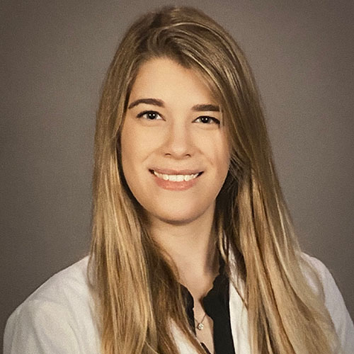
Samantha Glamann, DVM
Resident

Rachel Sullivan, VMD
Resident
Veterinary Support Staff
- Kimberly Bryan, R.T. (R)(M)
- Rebecca Bauer, R.T (R) (CT)
- Laurie Lyn Burton, R.T. (R)
- Samantha Jackson, R.T. (R)
- Chuck Lemmond, B.S. R.T. (R) (T)
- Kingslea Magana, Ultrasound Assistant
- Thea Partin, CNMT, RT(N), PET, NCT, NMTCB(CT), LVT
- Tatum Ramsey, R.T. (R)
- Jeanie Roberts, Ultrasound Assistant

